|
|||||||||||||||||
|
Differential diagnostics of oxyphilic lesions - table 1
|
|||||||||||||||||
|
|||||||||||||||||
|
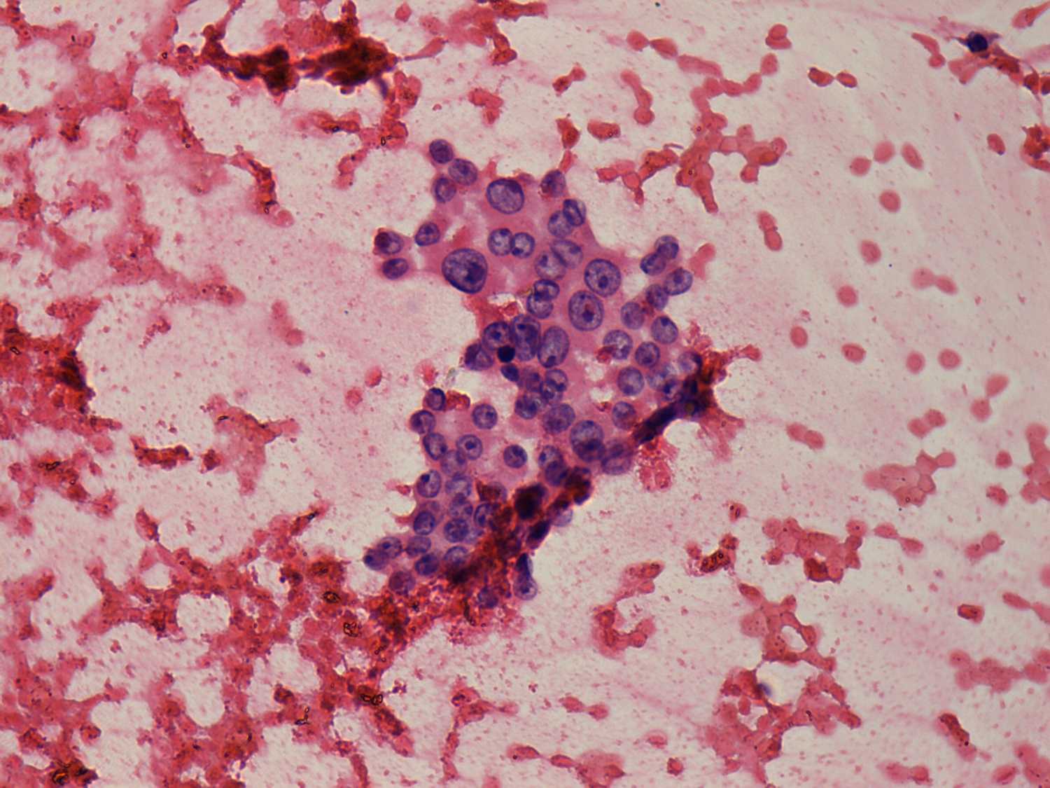
Oxyphilic ells are predominantly found in small cohesive groups. In the left case microfollicles can be seen, too and nuclei contain prominent nucleoli. On this presentation the left case may be either an oxyphilic tumor or a hyperplastic nodule. Taking the ultrasound presentation into account deeply alters our opinion. The nodule in the left case presents neither a halo nor a perinodular lood flow, therefore the chance for a follicular type tumor is negligible. |
|||||||||||||||||