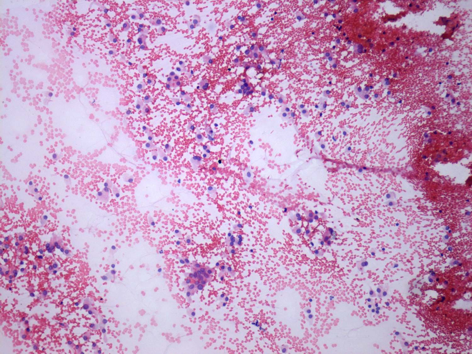
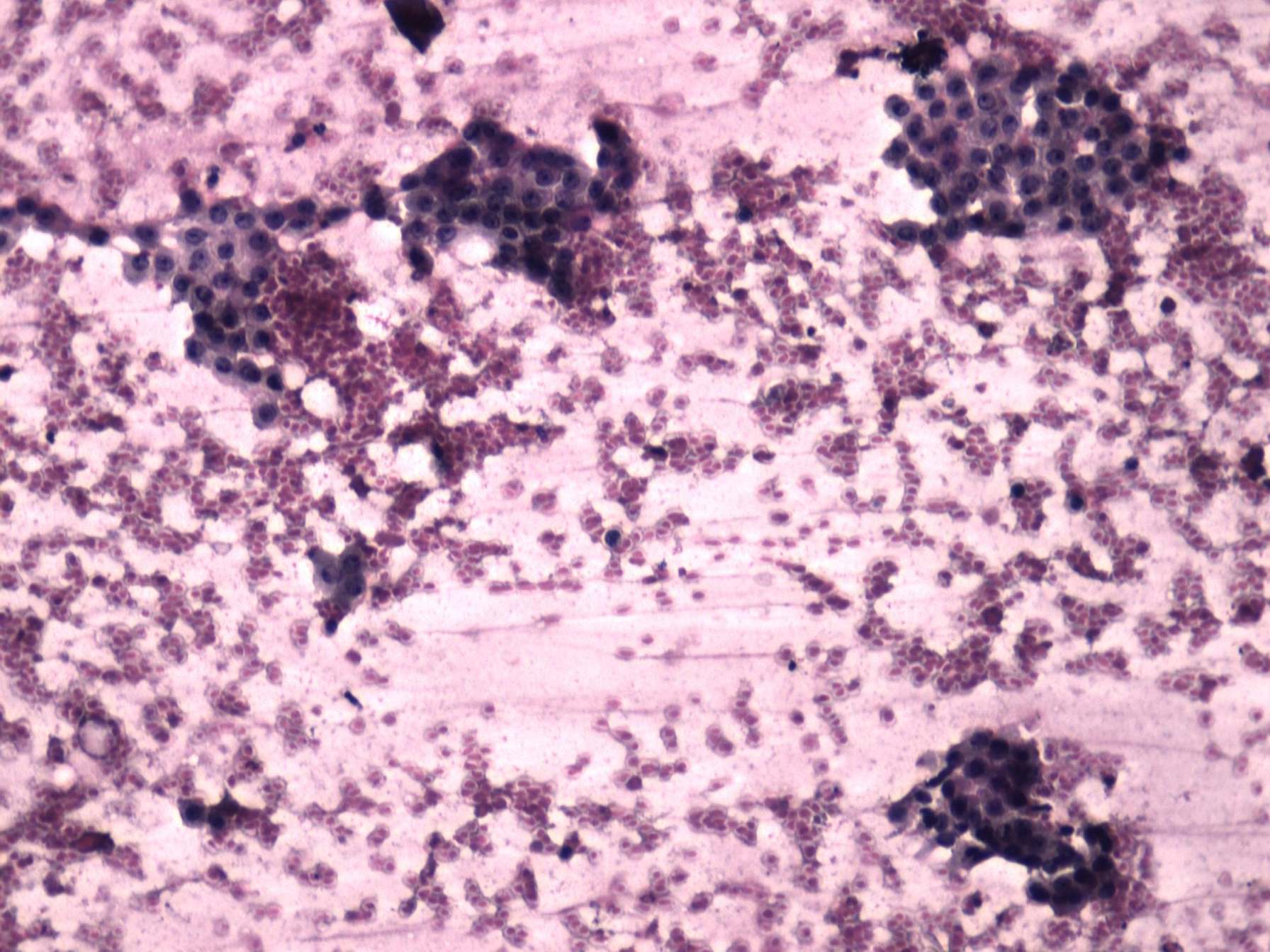
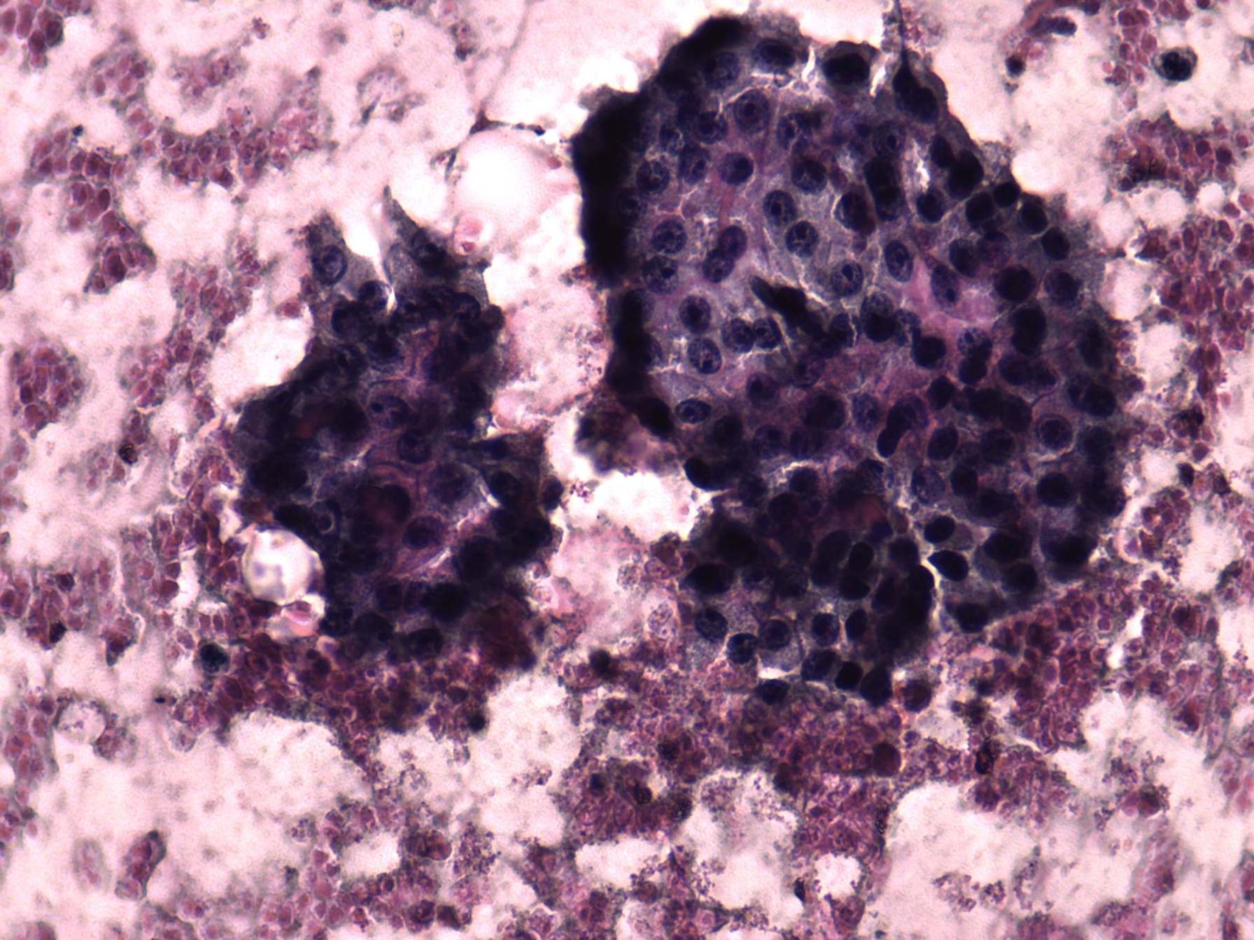
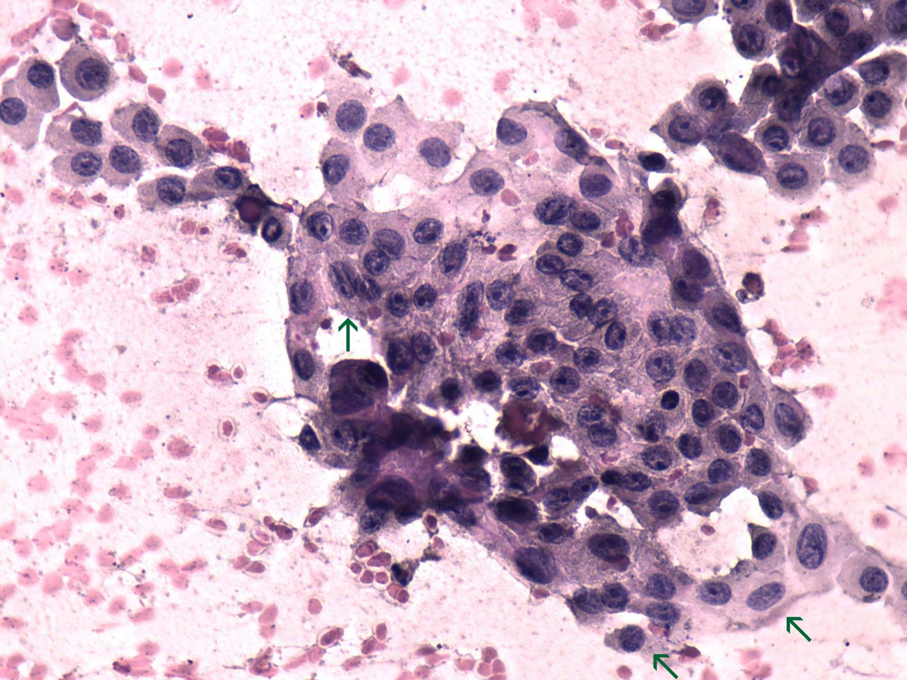
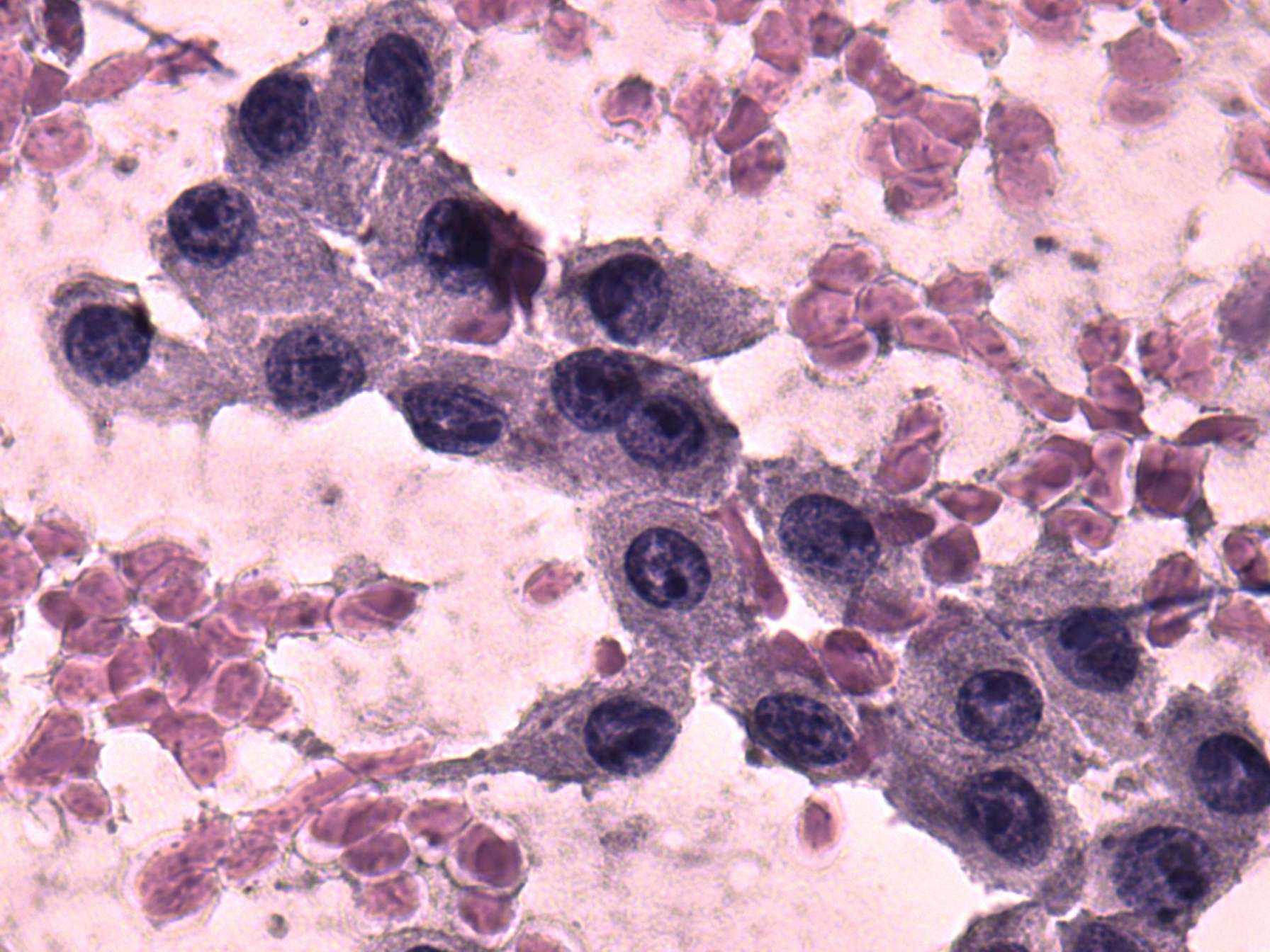
The hyperplastic nodule is characterized by the presence of monolayered sheets with only minimal degree of dissociation. The presence of prominent nucleoli increased the likelihood of tumor. In the event of the right case, isolated cells and microfollicles predominated the smear. Intranuclear grooves were found in both cases. This finding has a limited significance in metaplastic cells. Taking the cytological properties into account the risk of an oxyphilic tumor was relatively low in the left while high in the right case.
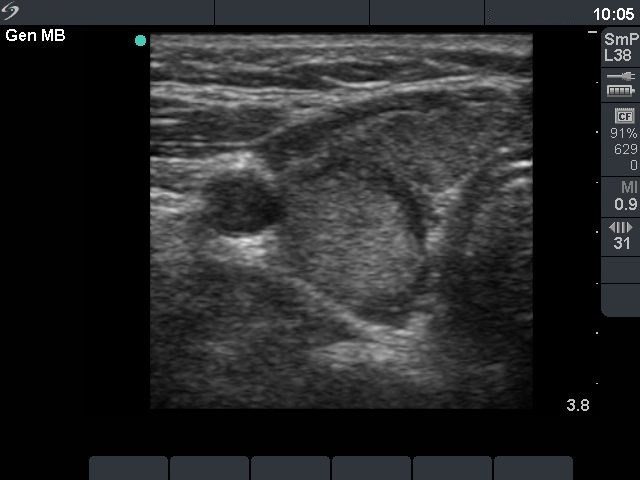
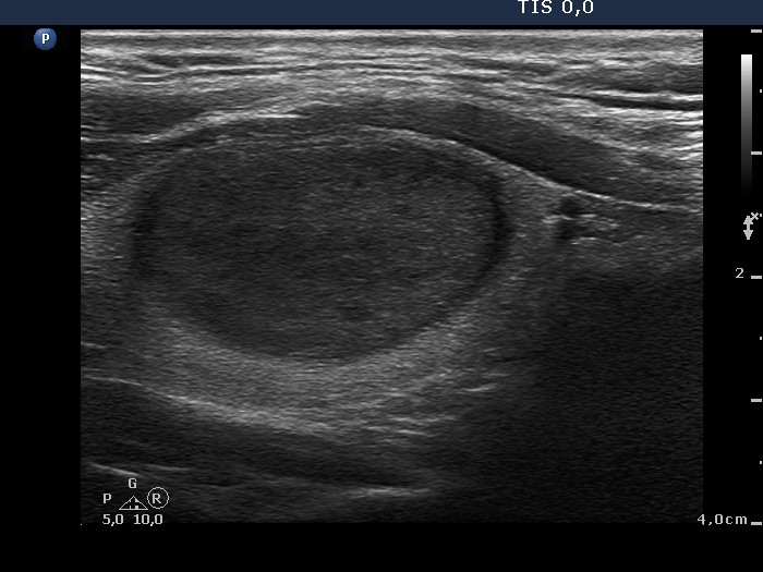
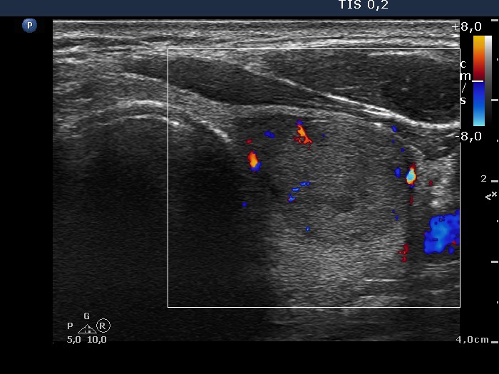
The ultrasound presentation decided the issue in the right case: a large solitary nodule presenting halo sign and signs of perinodular is by all probability a follicular tumor. The interpretation of the ultrasound pattern is more difficult in the left patient. She was operated on benign hyperplastic nodules for 18 years. The presence of discrete nodule-like lesions in an operated thyroid is a frequent finding. This lesion was irregular in shape which decreased the likelihood of a follicular tumor. The hypoechogenic rim differs from a usual halo, it probably corresponds to a thick connective tissue. The lesion is echonormal which would be a very unusual finding in a malignant tumor. Taking the cytological and ultrasound properties into account, our diagnosis was
benign recurrent nodule with the risk of malignancy less than 1:100.