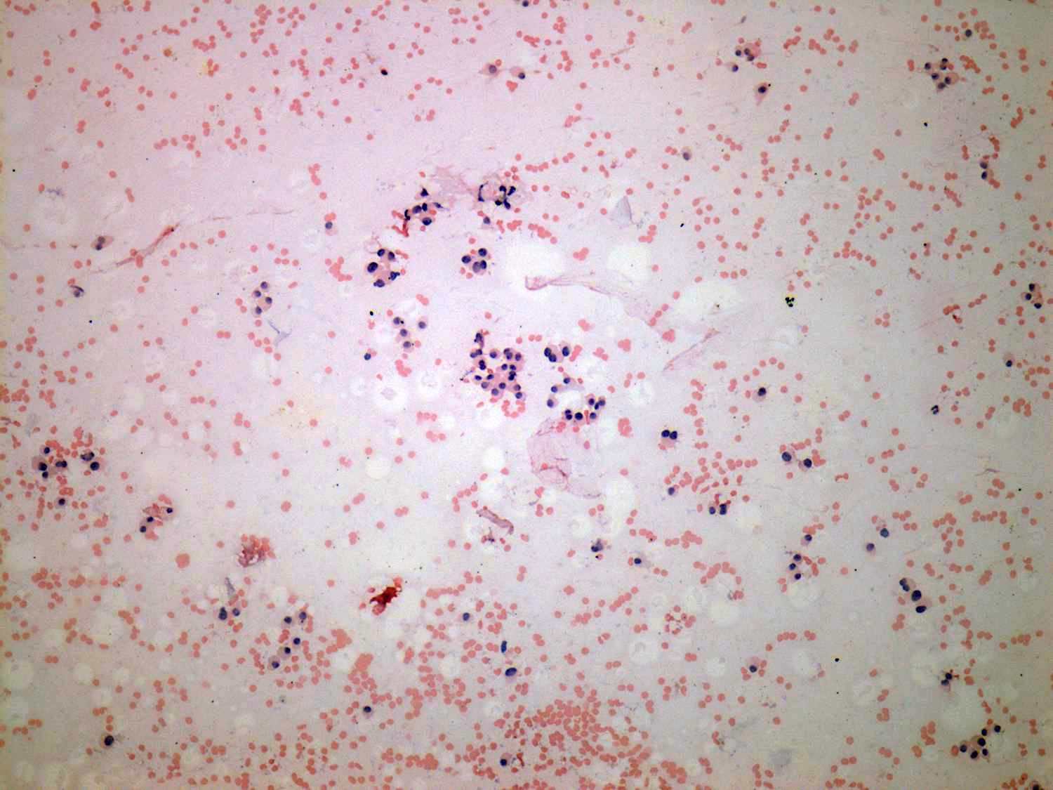
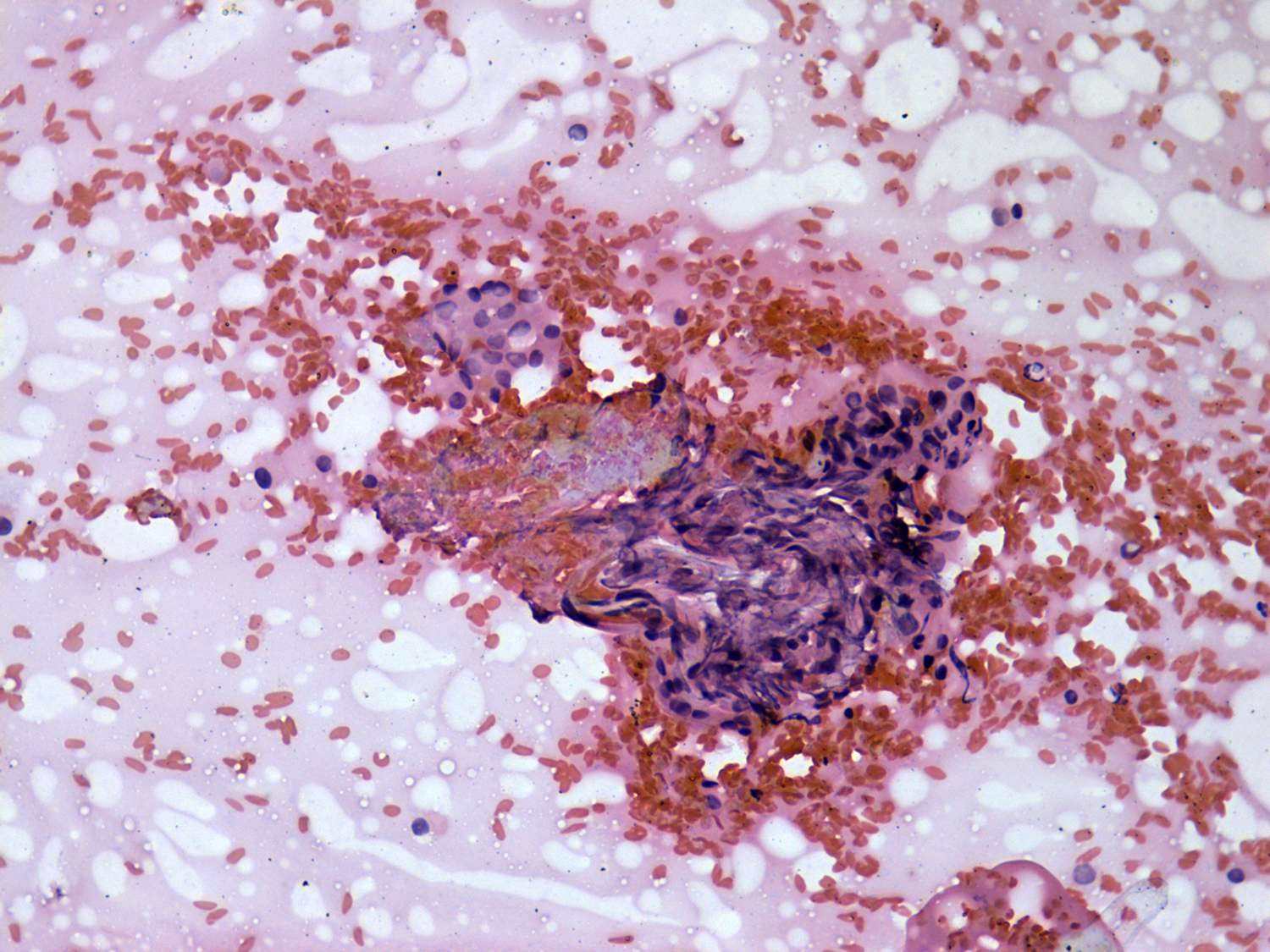
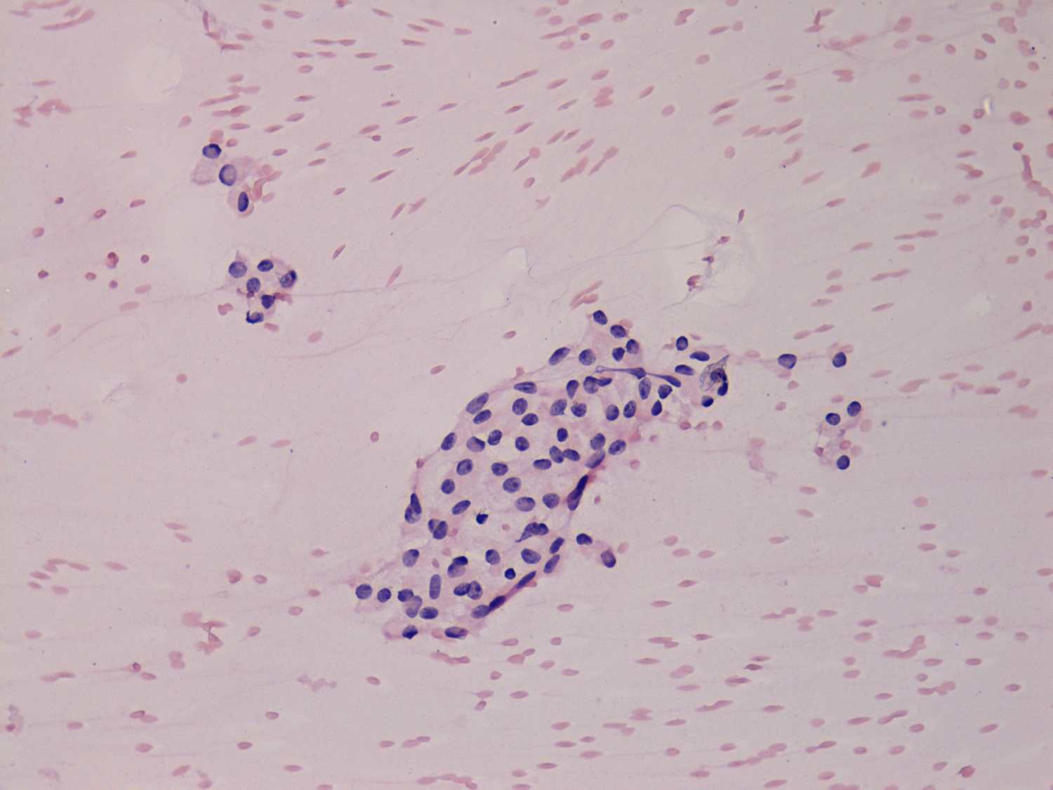
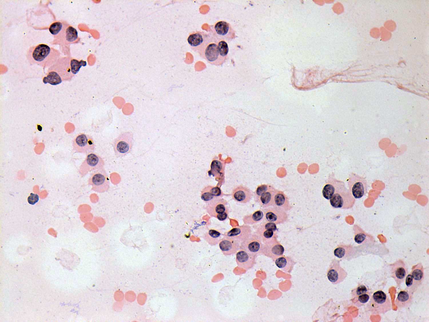
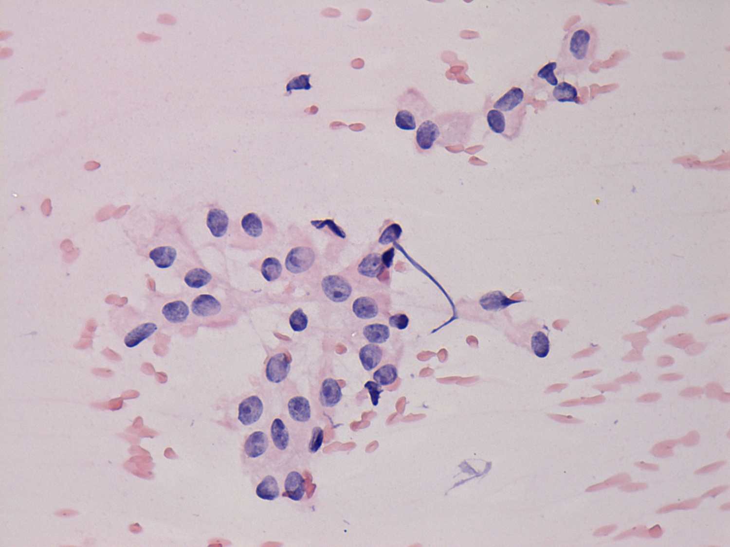
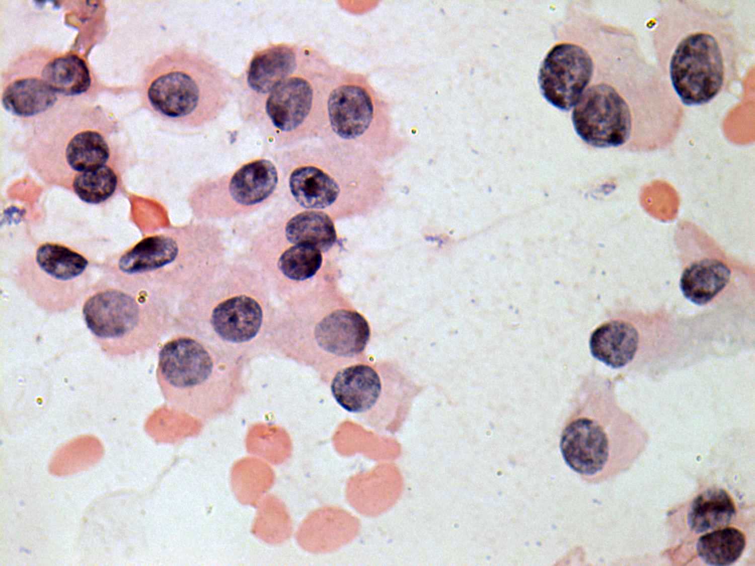
There is no difference in the cytological appearance of the two cases: mostly isolated oxyphilic cells are found on the smear. The left case is more suspicious of oxyphilic tumor because of the presence of prominent nucleoli. The lack of intranuclear inclusions and grooves makes the possibility of a papillary carcinoma very unlikely.
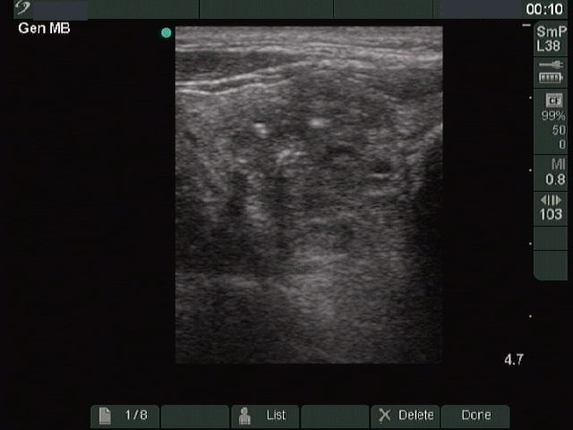
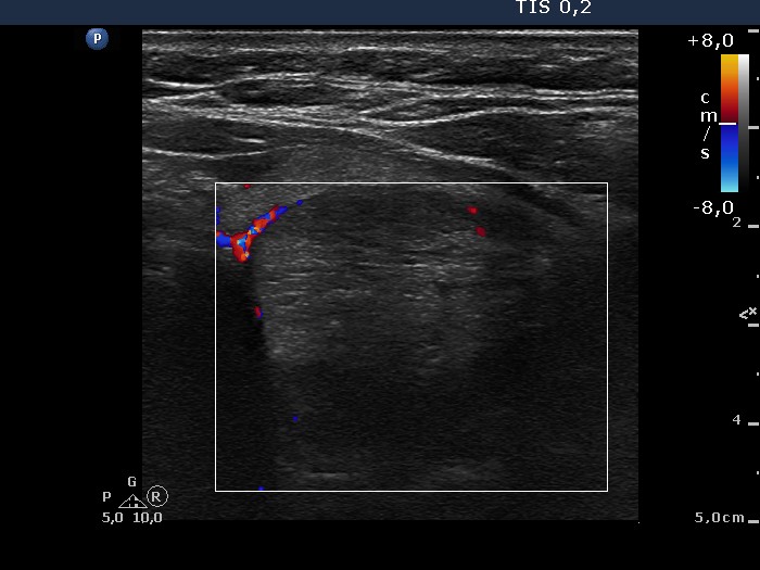
If we take the sonographic presentations into account, then the risk of an oxyphilic variant of a follicular tumor is unlikely in the event of the left case while very likely in the event of the right patient, because the former lacks while the latter presents halo sign and perinodular blood flow.