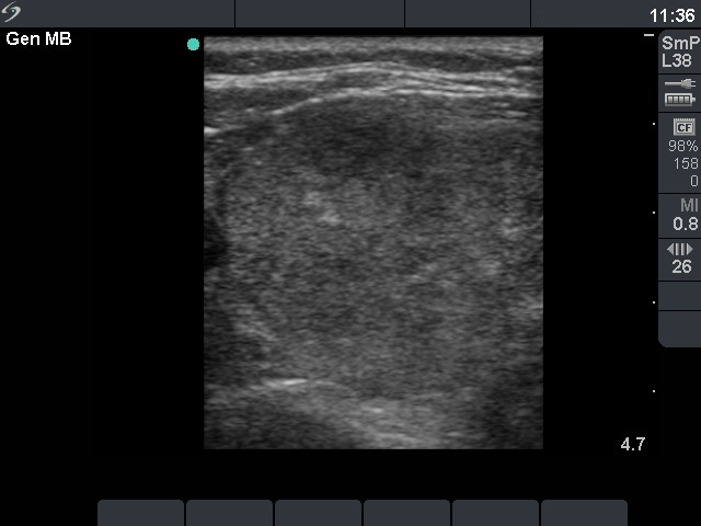
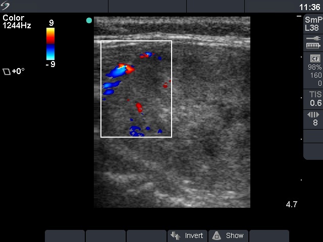
The cytological presentations of these cases are practically identical: oxyphilic cells are located predominantly in small cohesive groups, they lack prominent nucleoli, inclusions and grooves. Therefore the possibility of a papillary carcinoma is very low. A hyperplastic nodule and an oxyphilic variant of a follicular tumor have to be considered.
If we take the ultrasound pattern into account the risk of a tumor differs significantly: the hyperplastic nodule presents neither a halo nor a perinodular blood flow, while the oxyphilic adenoma does the latter.