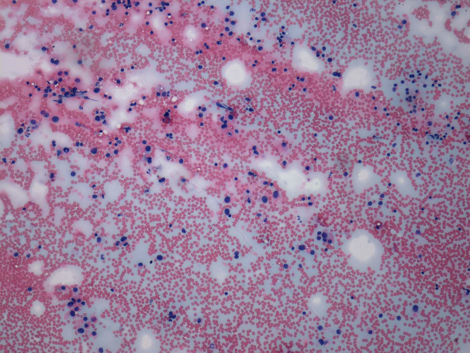
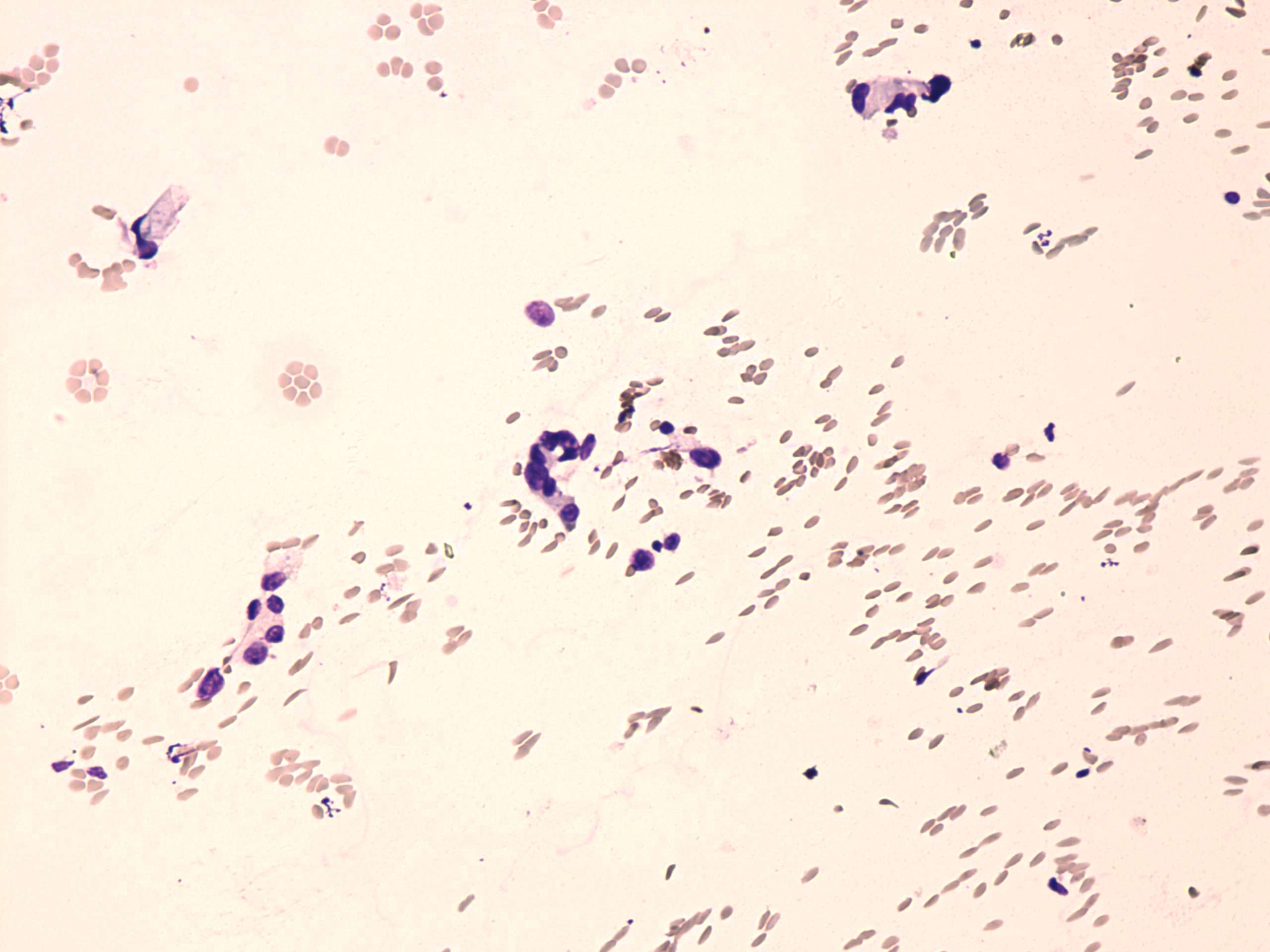
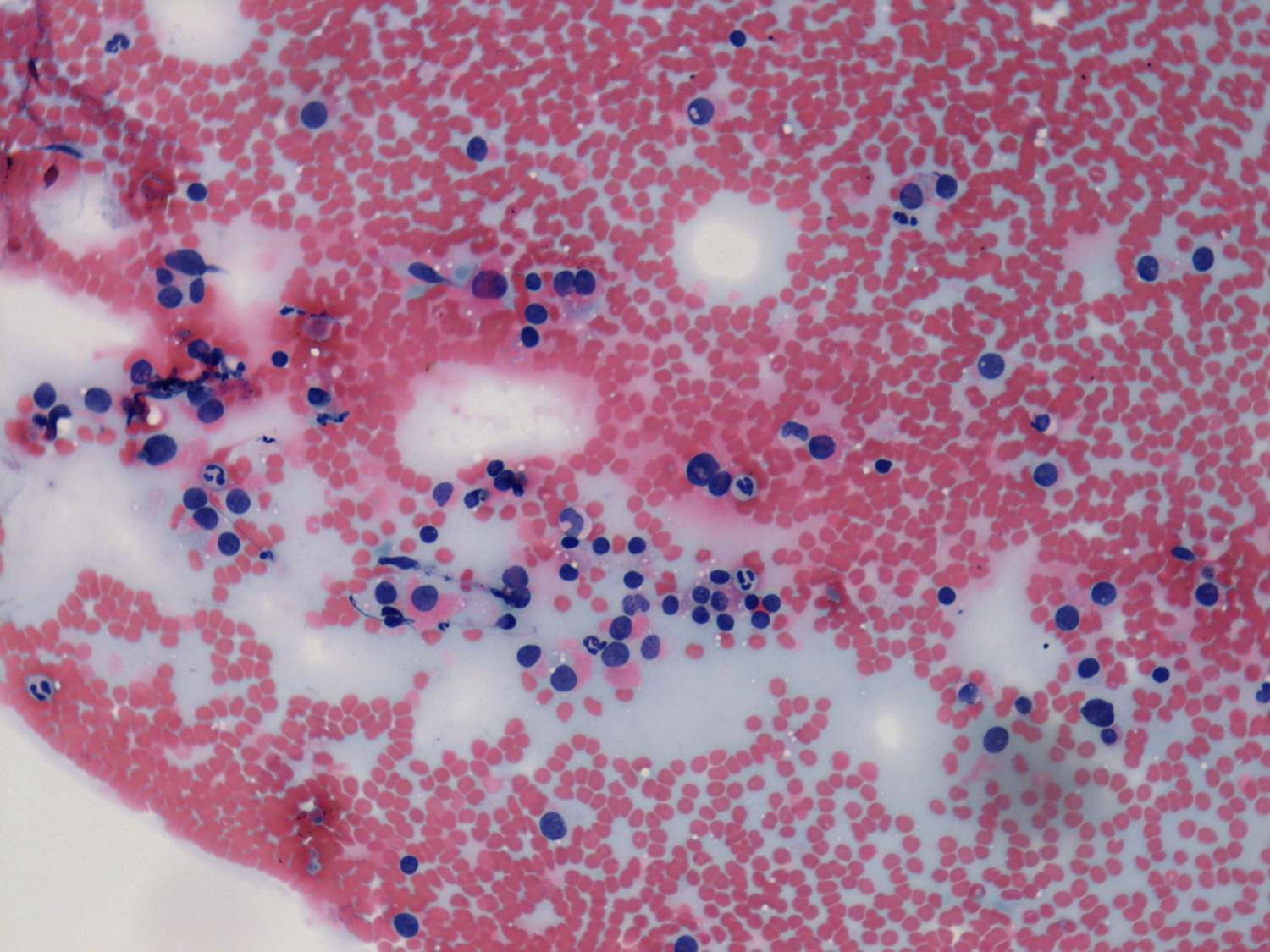
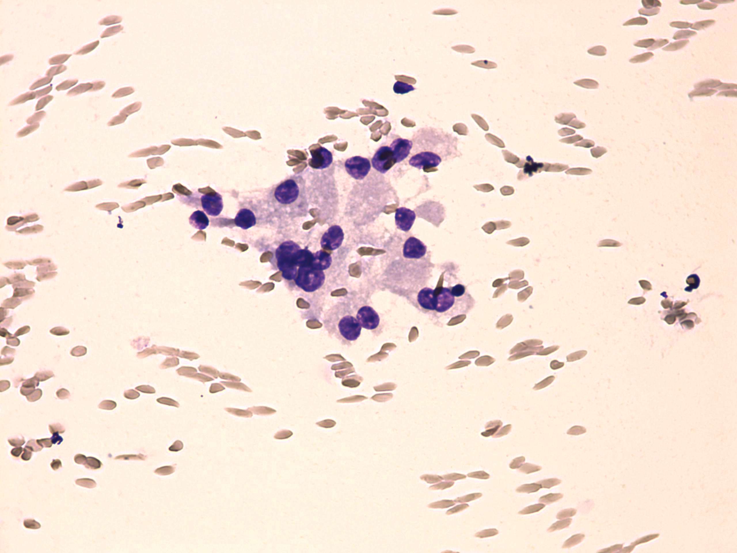
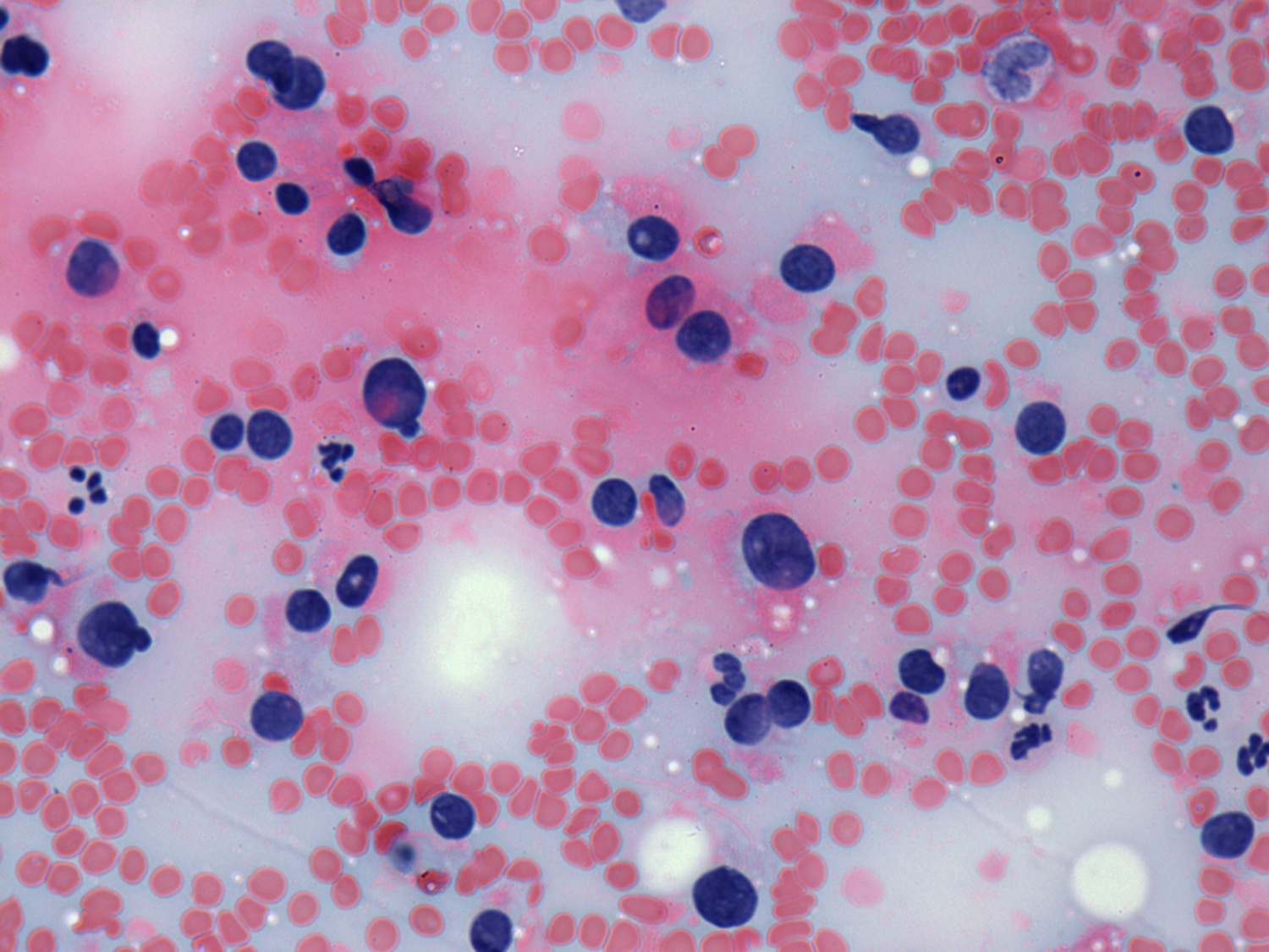
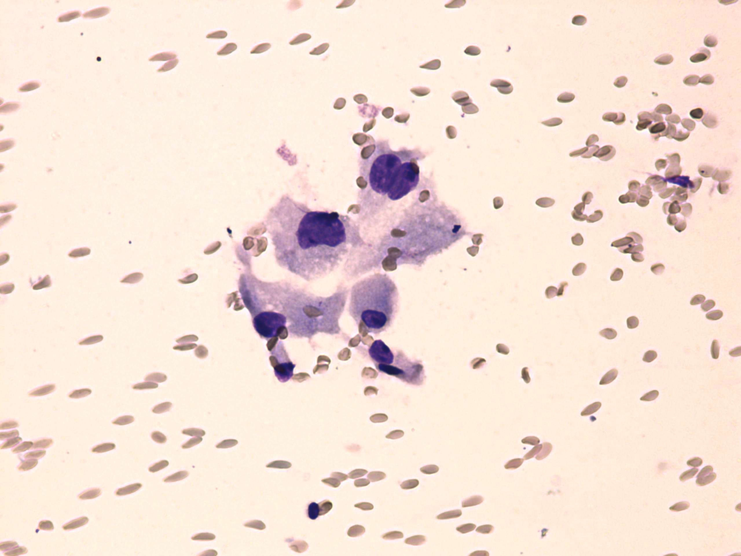
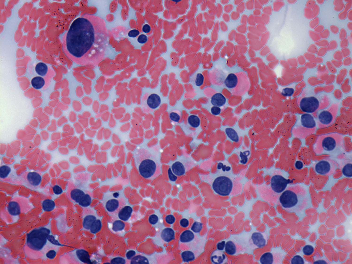
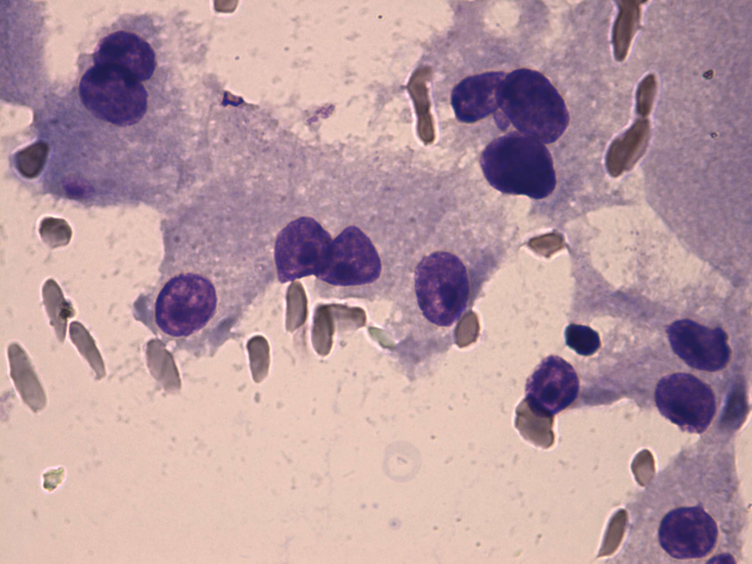
Although both cases present a lymphoepitheloid cell population, there are only few lymphocytes in the left while many lymphoid elements in the right case. Isolated atypical and even pleomorphic cells predominate the smears. The nuclei contain prominent nucleoli in the left case. The cytological pattern itself is very suspicious for a Hürthle-cell tumor in the left case while in the right one Hashimoto's thyroiditis and oxyphilic tumor are involved in the differential diagnostics.
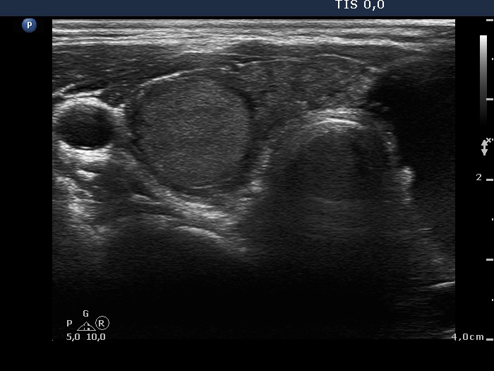
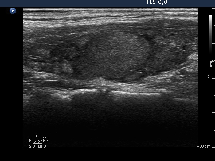
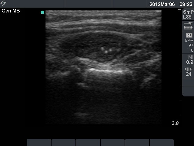
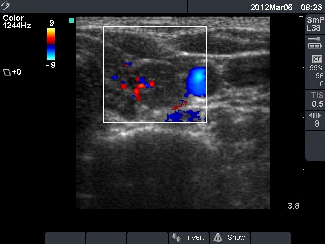
The ultrasound presentations fundamentally change the likelihood of certain diseases. There were several identical echonormal lesions in the left case which corresponded the so-called micronodular from of Hashimoto's thyroiditis, while there was a hypoechogenic lesion divided by connective tissue form the identically hypoechogenic thyroid. This discrete area contained microcalcifications and presented a type 3 vascular pattern.
As regards the right case, the cytological pattern practically disclosed the possibility of a papillary carcinoma while the ultrasound pattern did that of a follicular type tumor. Although the latter could not be excluded on the ultrasound presentation, the risk is low.