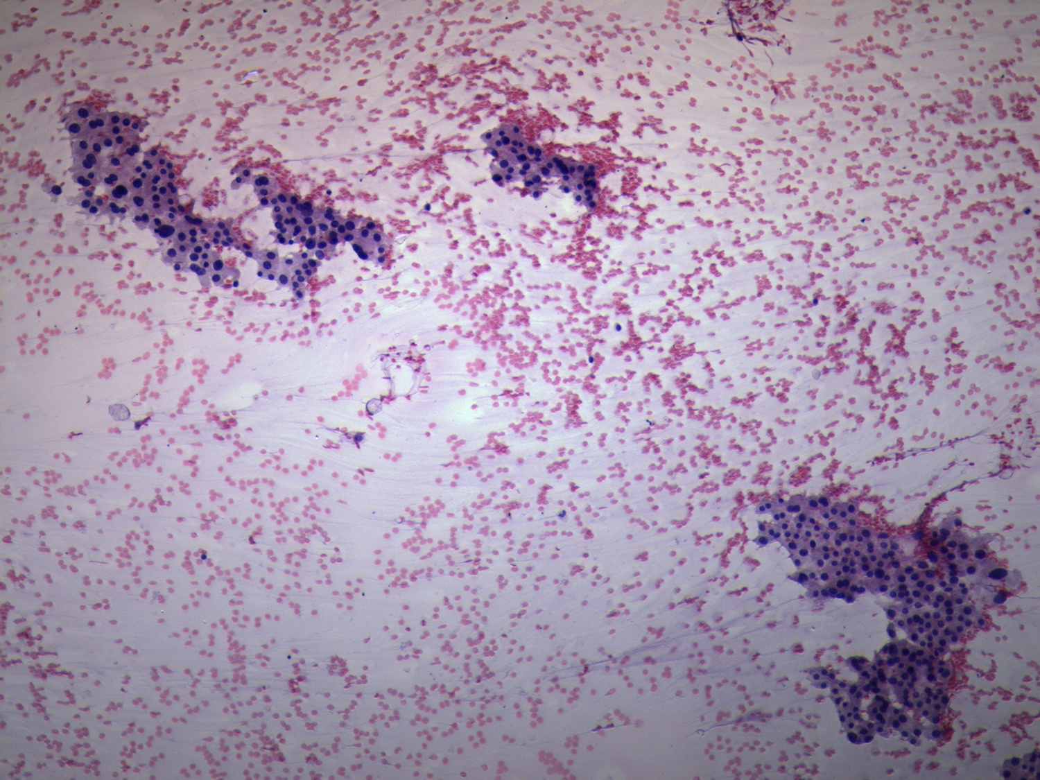
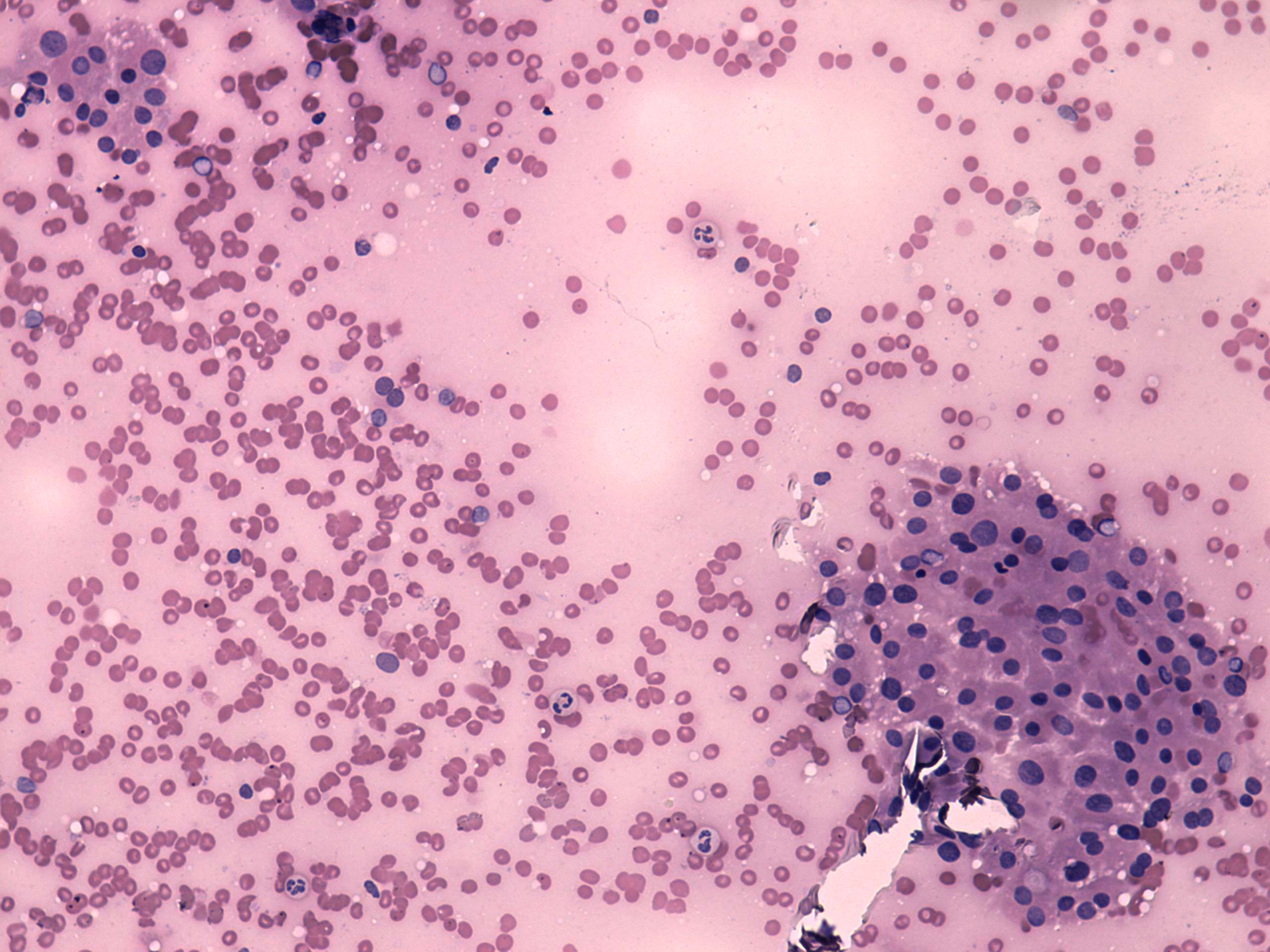
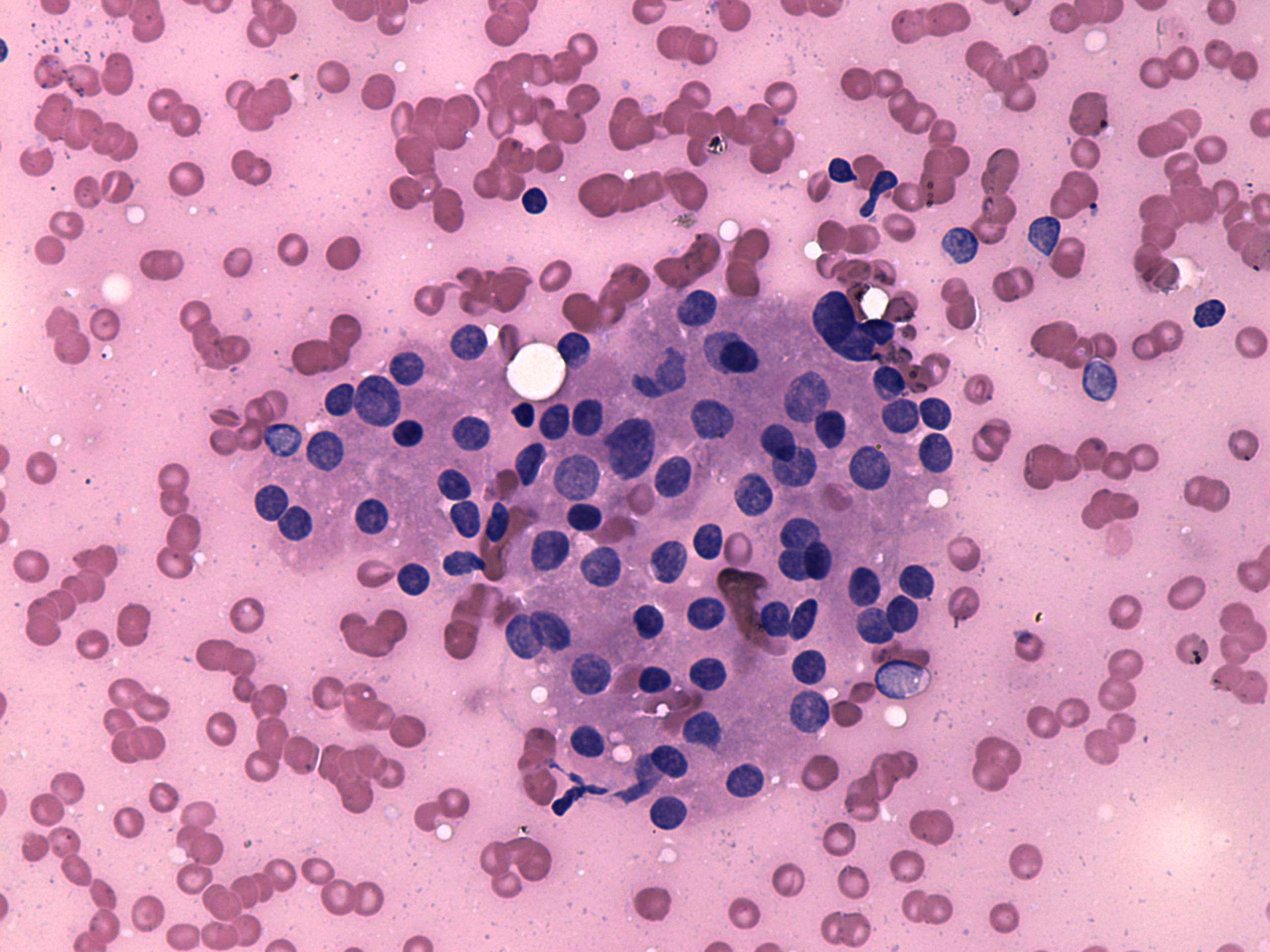
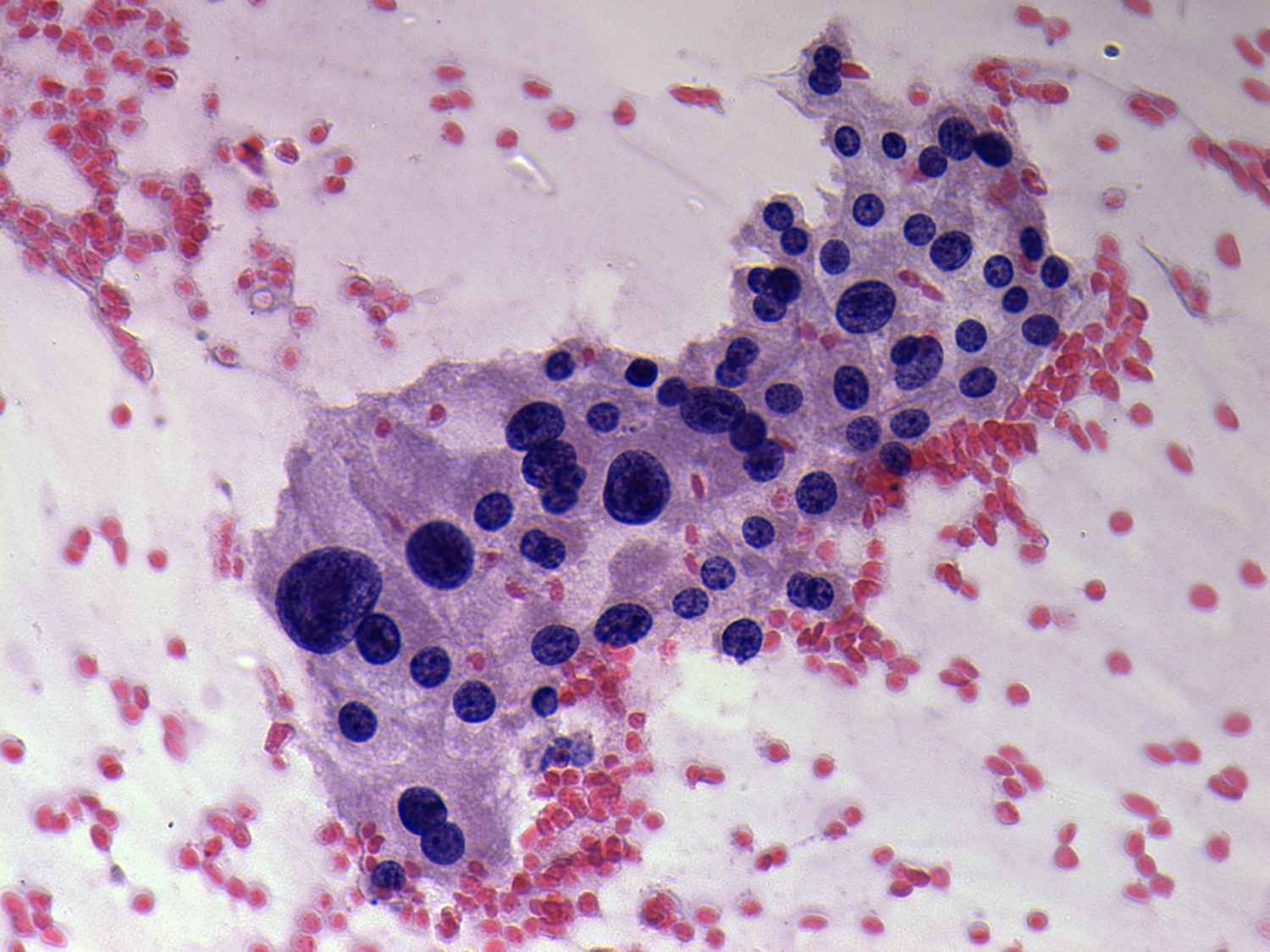
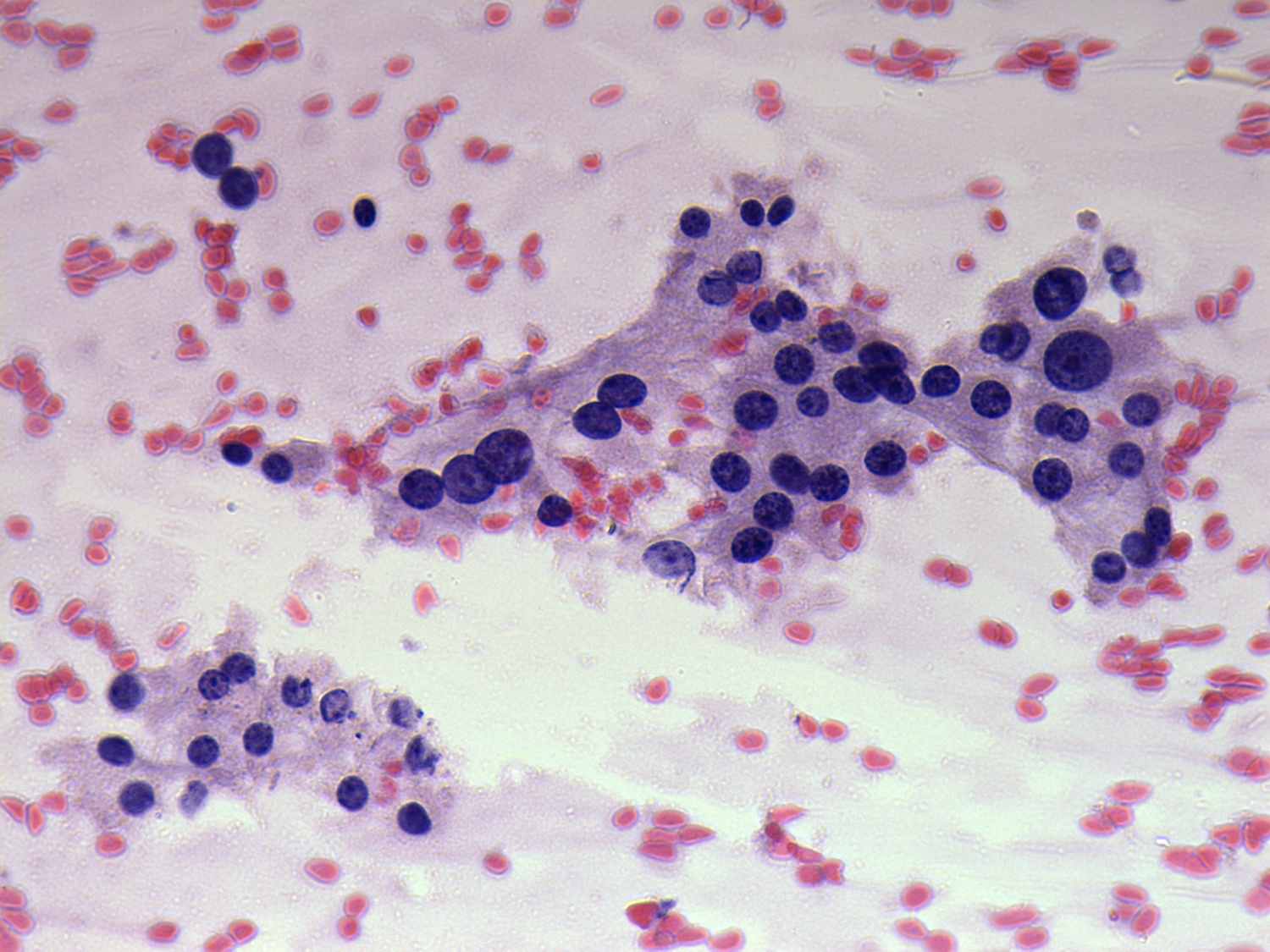
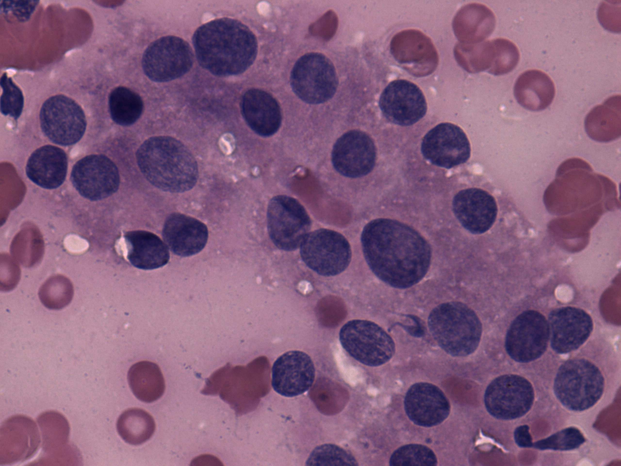
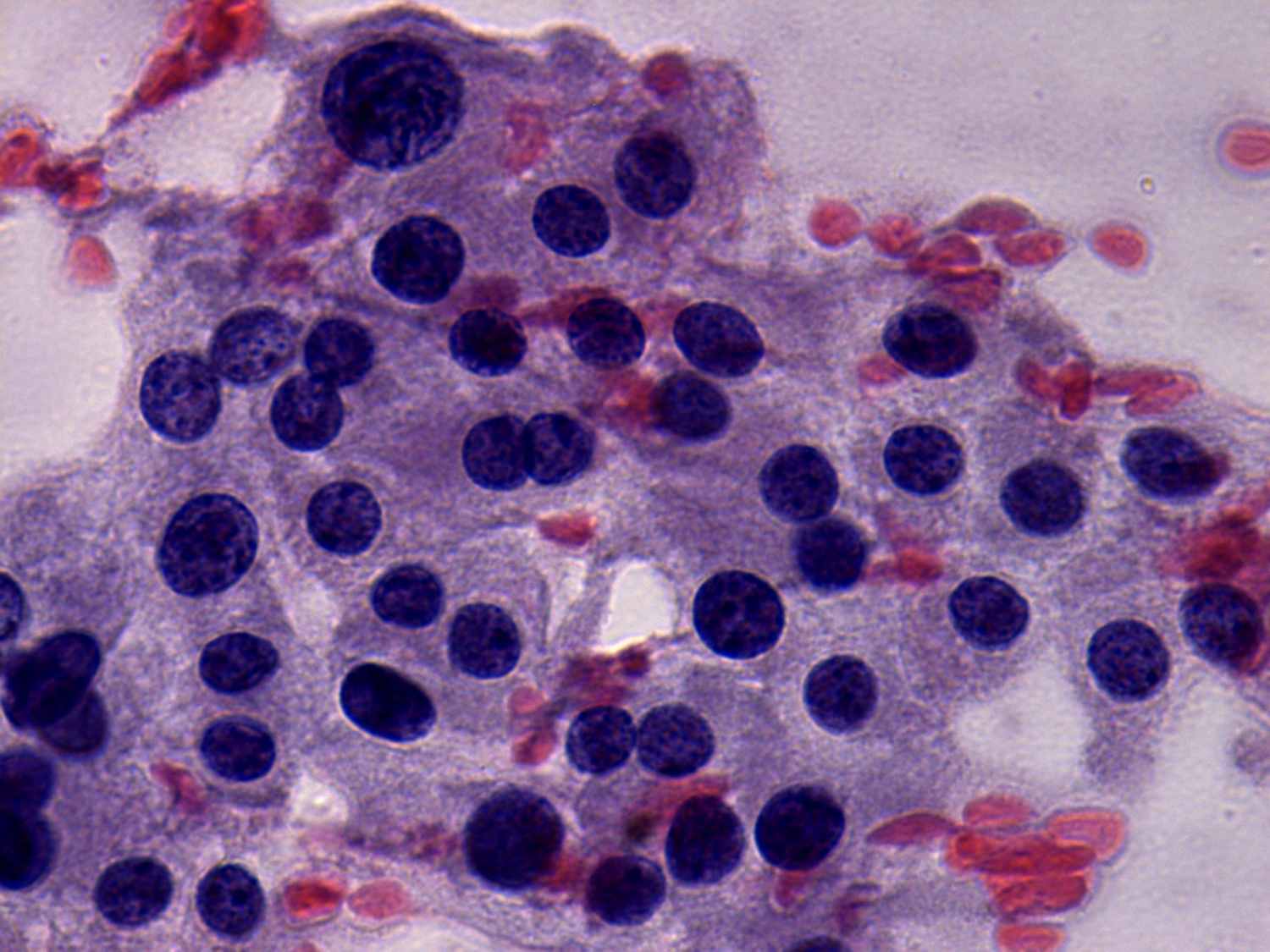
Both smears presented Hürthle-cell proliferation. There were a few lymphocytes in the left smear. Oxyphilic cells are located in small groups with relatively low tendency of cells to dissociate. A benign hyperplastic nodule, an oxyphilic variant of a follicular tumor, and Hashimoto's thyroiditis are involved in the differential diagnostic in order of probability.
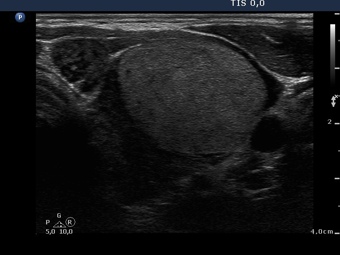
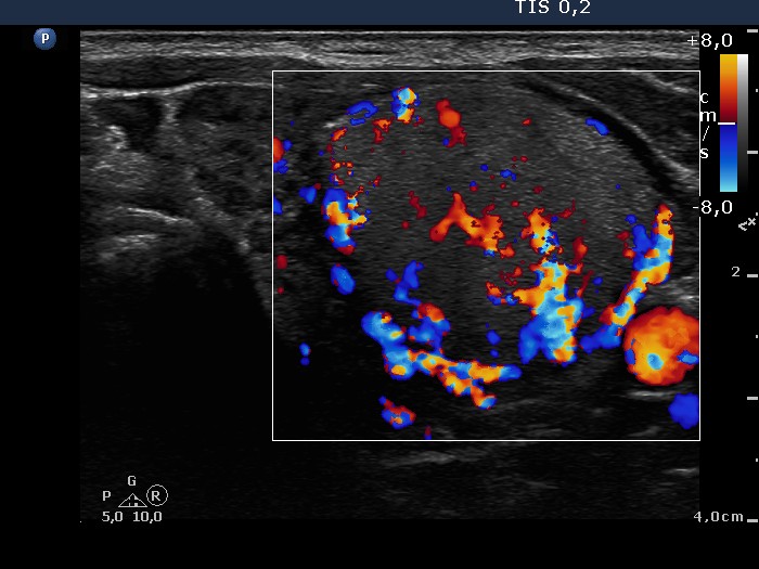
The ultrasound pattern changes the order of probability: there was a large solitary nodule presenting halo sign and perinodular blood flow in the left case which makes an oxyphilic variant of a follicular tumor the most likely diagnosis.
As regards the left case, based on the presence of multiple hypoechogenic lesions, Hashimoto's thyroiditis seems to be the most likely diagnosis. In fact, histopathology disclosed Hashimoto's thyroiditis without any nodule.