|
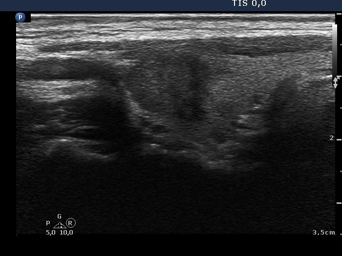
Transverse scan |
Longitudinal scan |
 |
 |
 |
 |
The nodule presents the taller-than-wide shape on transverse scan because the depth (antero-posterior diameter) clearly exceeds the width (latero-medial diameter) of the nodule. On the hand longer-than-wide shape is not present on longitudinal scan because the depth (antero-posterior diameter) is lower than the length (the diameter measured from the upper to the lower pole of the nodule). The depth is signed with white, the width is with red while the length is with yellow arrows. The nodule shows another suspicious sign, irregular borders. Note the spiculations on the medial and dorsal part of the nodule (blue arrows).
|
| |
|
Papillary carcinoma (histology) - case 218
|
Transverse scan |
Longitudinal scan |
 |
 |
 |
 |
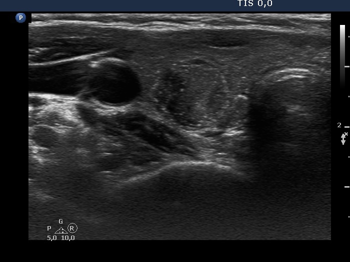
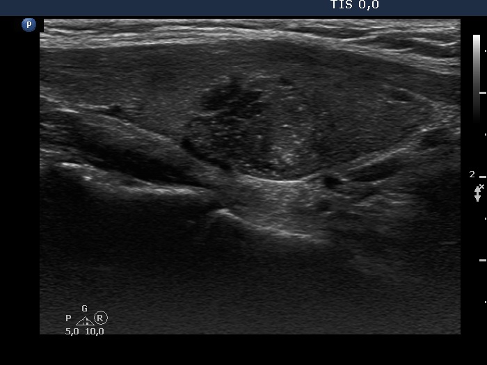
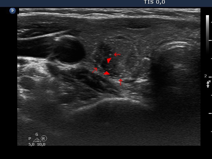
It is not evident whether the nodule should be regarded as deeply hypoechoic or not because the proportion of deeply hypoechoic parts are roughly equal to non-deeply hypoechoic portion. How, the nodule presents microcalcifications.
The echogenic granules are not as bright as in a usual case, however great proportion of them are microcalcifications (arrows). It means that the nodule is an EU-TIRADS 5 lesion.
|
| |
Metastasis of a laryngeal adenocancer to the thyroid (histology) - case 378
|
Right lobe |
Left lobe |
 |
 |
 |
 |
Both the nodule in the right and in the left lobe are clearly EU-TIRADS 5 lesions. The right due to the irregular margins, the left due to the deeply hypoechogenicity and nonparallel orientation.
|
| |
|
|
Transvers scans |
Longitudinal scans |
 |
 |
 |
 |
The upper nodule has microcalcifications while the lower is deeply hypoechoic. Both have irregular borders. These are clearly an EU-TIRADS 5 lesions.
|
| |
|
| |
|
| |
|
| |
|
| |
|
| |
| |
| |
|
