
Papillary carcinoma - Case 30. (ultrasonographic picture 5)
|
|

|
|
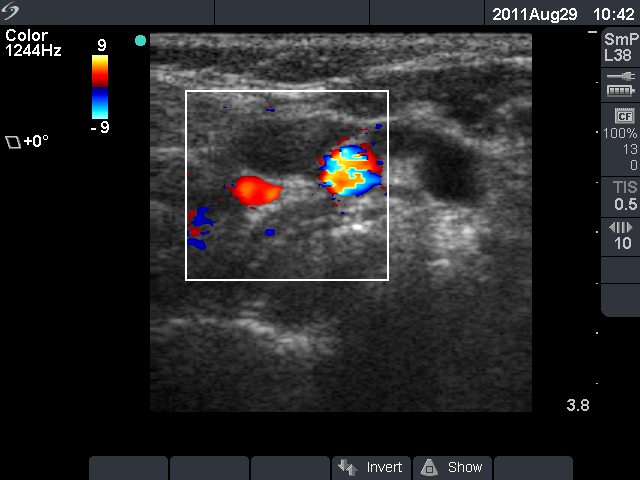
Above the left lobe, horizontal scan, color Doppler mode. The Doppler mode proves that one of the hypoechogenic lesions is not a vessel but a lymph node.