
|
|
Benign nodular hyperplasia - Case 44.
|
|


Clinical presentation: a 22-year-old woman with a nodule discovered on a routine examination. The patient had no complaints. A nodule was palpable in the right lobe.
Functional state: euthyroidism (TSH 1.29 mIU/L, FT4 21.8 pM/L).
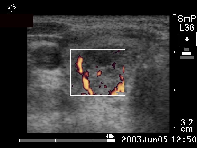
Ultrasonography: a solitary hypoechogenic nodule with central cystic degeneration.
Cytological report: a microfollicular pattern, no colloid in the background.
Cytological diagnosis: follicular tumor with less than the average risk for malignancy.
Scintigraphy disclosed an autonomously functioning nodule.
Suggestion: follow-up examinations with TSH determination every year, ultrasonography every three year.