Introduction - case 286 (ultrasonographic picture 4)

|

|

|
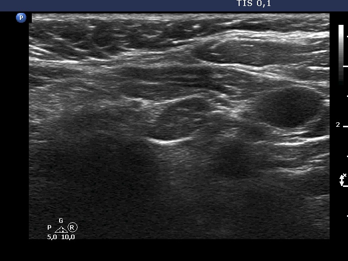
Lower pole of the left lobe, transverse scan. Here we can see a very hypoechoic mass.

Papillon Course
2024-25
Introductory courses
|
|
|
|
Lower pole of the left lobe, transverse scan. Here we can see a very hypoechoic mass.


