
|
|
Introduction - case 286
|
|


Clinical presentation: A 68-yr-old woman was referred for evaluation of a thyroid nodule detected on PET-CT scan which was performed on follow up of breast cancer.
Palpation: no abnormality.
Laboratory tests: TSH 2.12 mIU/L.
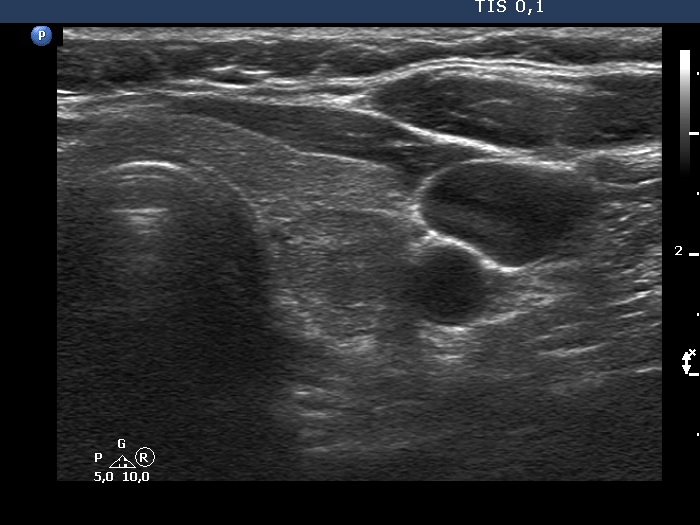
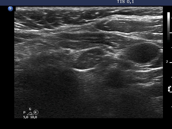
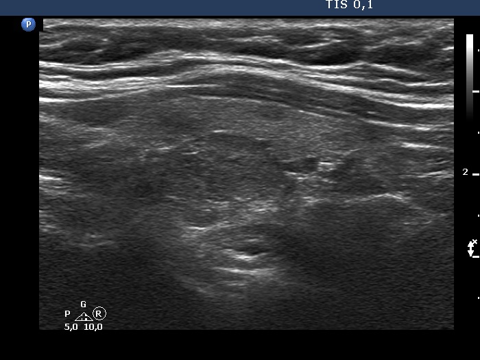
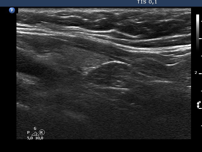
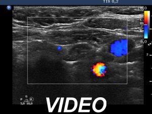
Ultrasonography. The thyroid was echonormal. According to the PET-positive lesion, there was a moderately hypoechoic nodule in the middle-dorsal part of the left lobe. The lesion presented with perinodular blood flow. In the transverse section, it appeared that there was another nodule in the lower pole of the lobe. At the same time, based on the longitudinal section, it was not an oval, nodule-like lesion. The mass proved to be longitudinally elongated in the caudal direction and was avascular.
FNA of the nodule in the middle part of the left lobe resulted in benign colloid goiter.
Comment.
-
An anatomical structure running perpendicular to the transducer looks round or oval, and it can look deceptively like a discrete lesion. If this occurs in the thyroid region, it can be mistaken for a nodule. The most common examples of this are the carotid artery, the jugular vein, and the esophagus, the latter usually appears on the dorsal surface of the left lobe of the thyroid gland. The key is always to examine the patient in two perpendicular planes. An examination parallel to the anatomical structure (in this case in the longitudinal section) shows that it is a tube-like, longitudinal structure. In this case, it was very likely a dorsal muscle.
-
A muscle used to be avascular on Doppler mode.