
Introduction - case 286 (ultrasonographic picture 8)
|
|

|
|
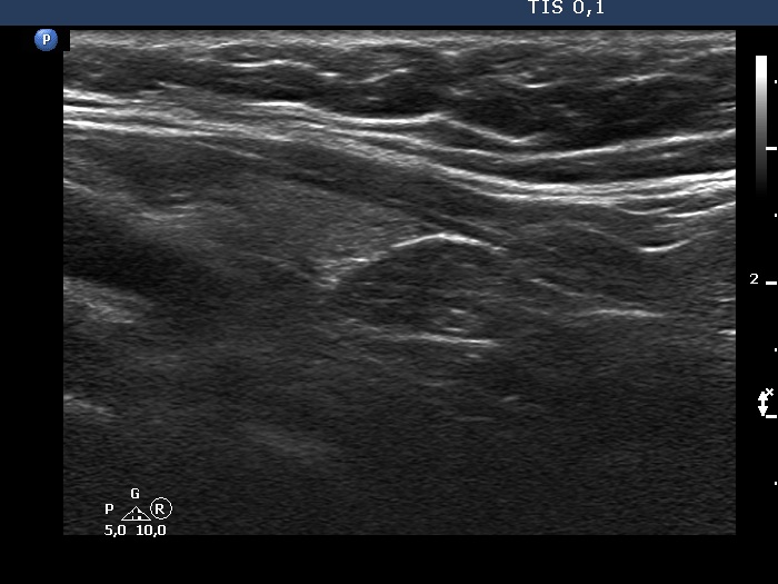
Lower part of the left lobe, another longitudinal scan - bit lower than the previous image. In this section, the lower pole cannot be defined, indeed the mass follows caudally (right in the image) to a muscle fiber.