
Introduction - case 286 (ultrasonographic picture 7)
|
|

|
|
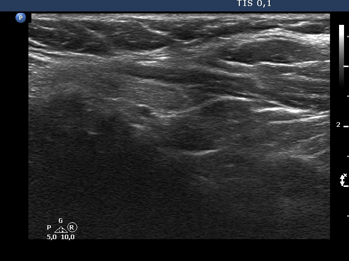
Lower part of the left lobe, another longitudinal scan - bit lower than the previous image. The nodule is more elongated and its lower pole is much less evident.