
|
|
Intranodular hyperechogenic figures - case 808
|
|

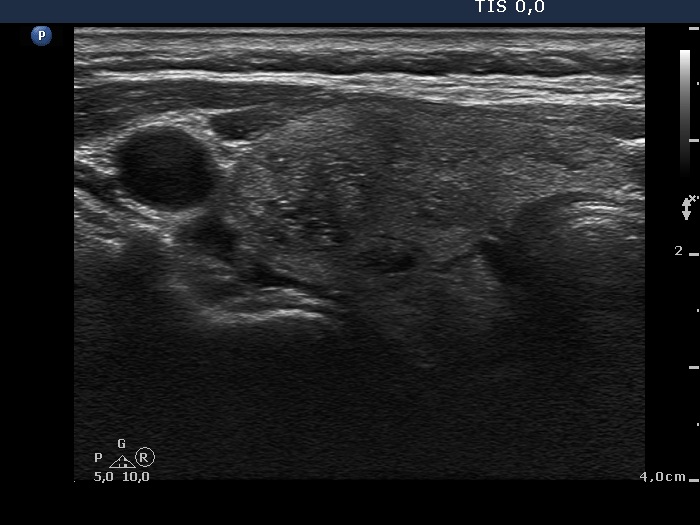


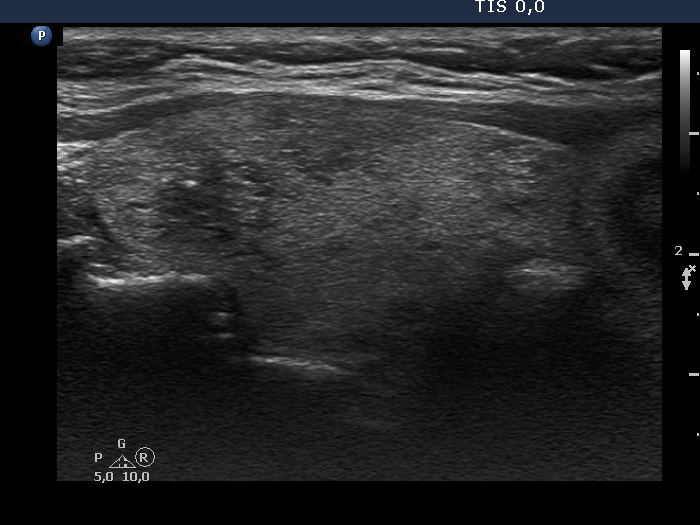

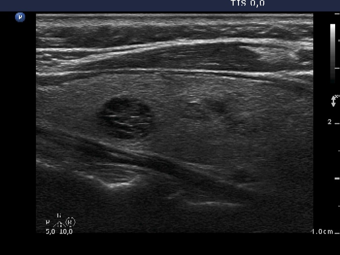
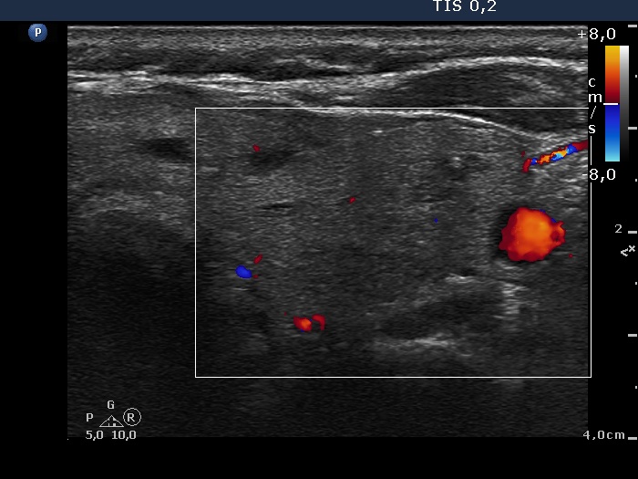
Left side of the isthmus


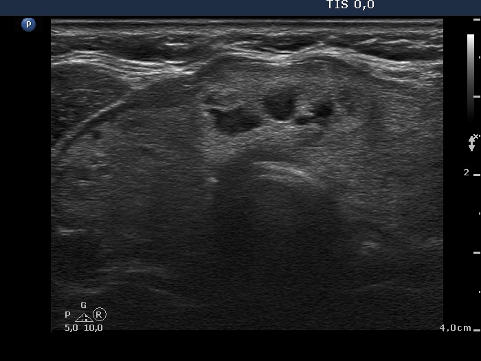


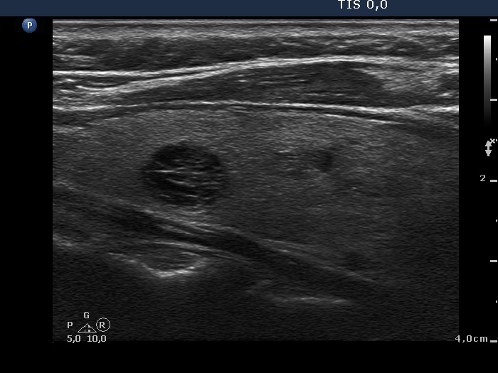
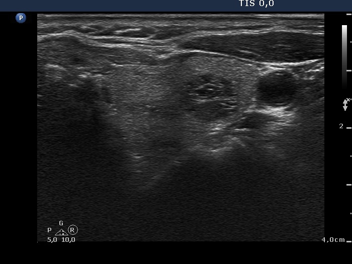
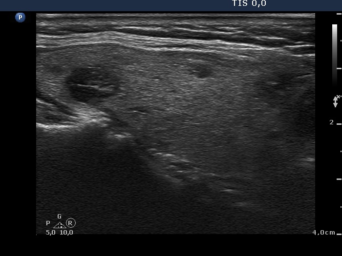
Left lobe


Note that in this case study the arrangement of the images differs from other case studies.
Clinical presentation: A 62-year-old woman was referred for an evaluation of nodular goiter detected by her GP on routine examination.
Palpation: Not firm nodules were palpated in the right lobe and in the isthmus.
Result of blood tests: euthyroidism (TSH 2.26 mIU/L).
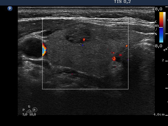
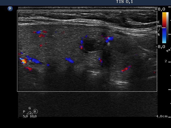
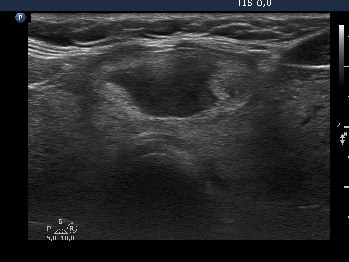
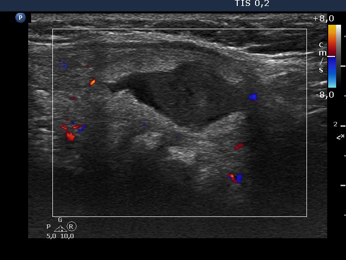
Ultrasonography: The thyroid was echonormal. There were several moderately hypoechogenic-cystic lesions in both lobes and a mixed echonormal-cystic lesion in the isthmus. The nodules showed various forms of intranodular figures.
Cytology was performed from two nodules, one in the right and the other one in the left lobe and resulted in benign lesions.
Summary of follow-up: neither the function nor the size and pattern of the nodules had changed over the follow-up except for the nodule in the isthmus which had changed in size at both follow-up occasions.
Comments.
-
The presentations of the nodules in the right and left lobe did not change over the 5 years of follow-up, moreover they remained almost the same.
-
It is worth analysing the solitary bright hyperechogenic granule in the right lobe. The presentation of this figure at the first and at the third examination was equivocal while at the intermediary examination it became evident that the figure is a comet-tail artifact.
- Most of the echogenic figures presented in the nodules are back wall figures.