
|
|
Thyroid cancers - case 1178
|
|

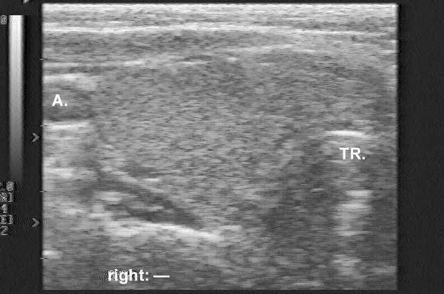


Clinical presentation: A 48-year-old woman who had been treated for Graves-Basedow's disease attended a follow-up examination. The patient had been examined 2 months earlier, when US was performed and revealed a nearly normal echo pattern without any discrete echo abnormality (see the first ultrasound picture). She was then well. Two weeks before the present examination, she felt dyspnea which progressed over the next 2 weeks.
Palpation: The thyroid was moderately enlarged. A hard mass could be palpated in the right thyroid bed.
Functional state: euthyroidism.
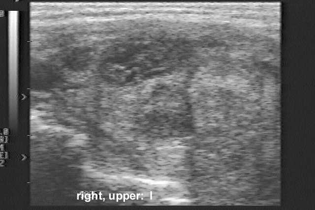
Ultrasonography revealed a hypoechogenic inhomogeneous nodule in the right lobe of the thyroid (the second US picture). The palpable mass was a hypoechoic lesion which was located just ventral to the right thyroid lobe.
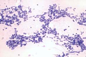
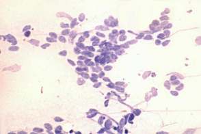
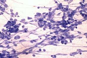
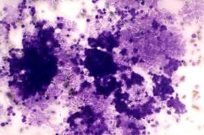
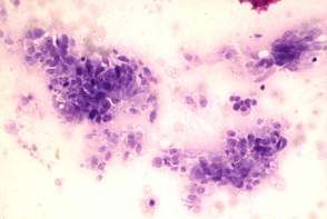
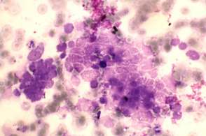
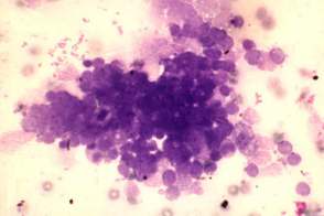
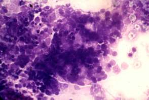
Cytology from both lesions resulted in small cell malignant tumor. Based on nuclear molding we raised the possibility of small cell lung cancer, which was confirmed by immunocytochemistry: the tumor cells did not express LCA but gave a positive reaction for neuron-specific enolase.
The patient was referred for a pulmonary evaluation. Bronchoscopy revealed a tumor in the lower lobe of the right lung.
Bronchoscopic brush cytology: small cell lung cancer.