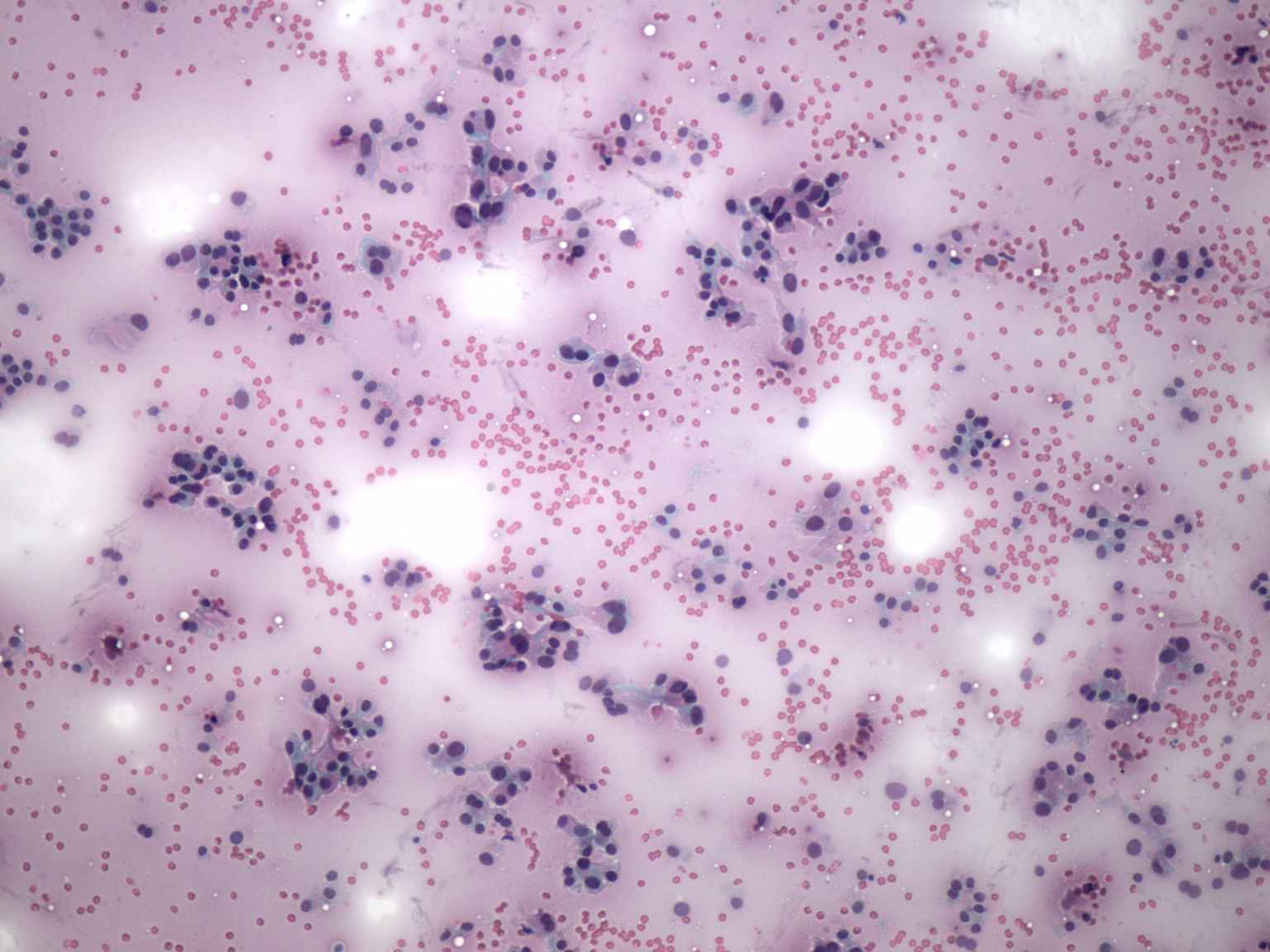
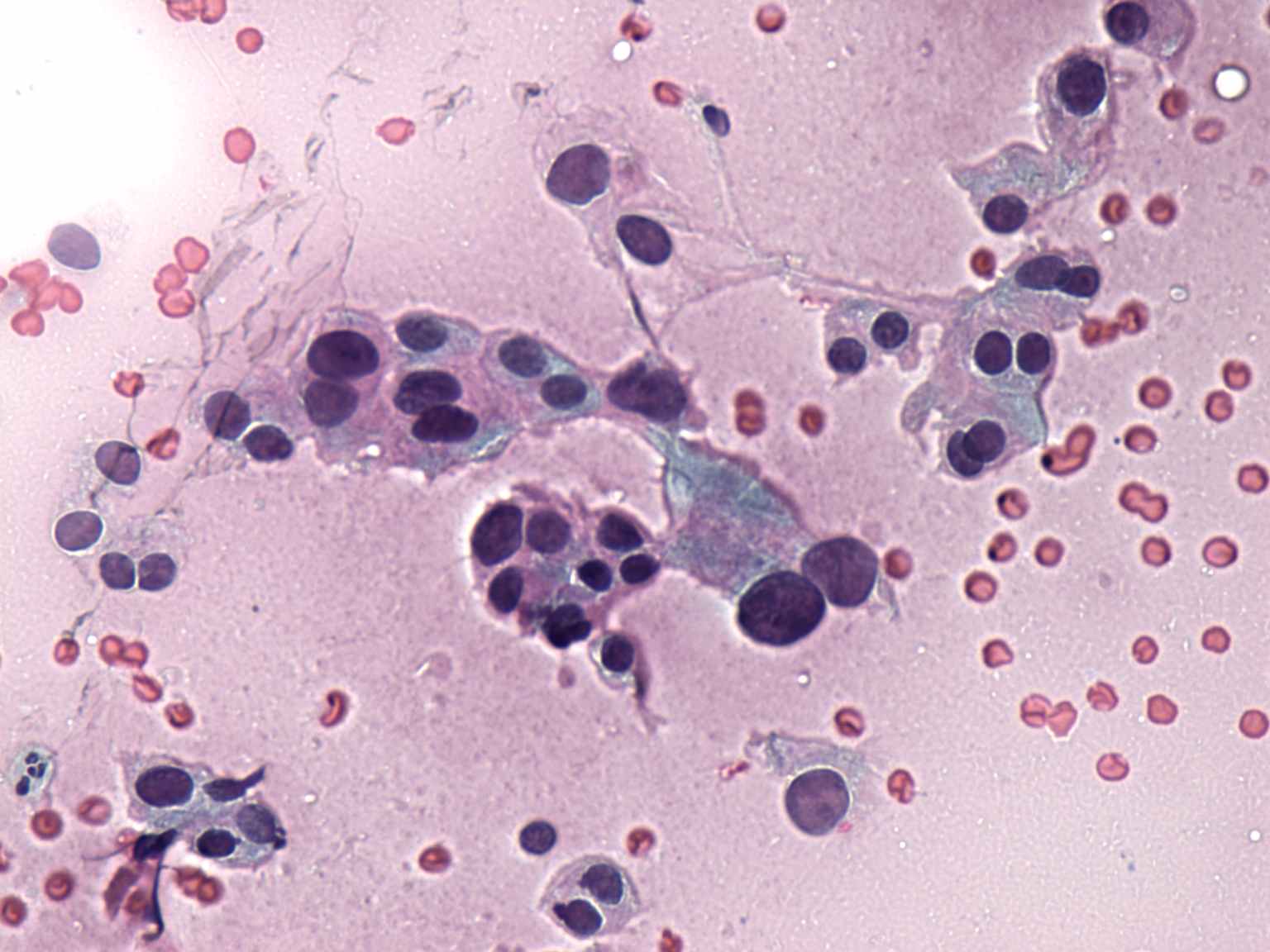
Compare the left and right images. Histologically verified
Hashimoto's thyroiditis is demonstrated on the left, while
histologically proven oxyphilic cancer on the right one. In the case
of the tumor, single cells predominate the smear, while in the case of
thyroiditis follicular cells are found in more cohesive structures. An
even more important difference can be observed analyzing the occurrence
of prominent nucleoli. Most oxyphilic tumors exhibit prominent
nucleoli, while in the case of Hashimoto's thyroiditis, prominent
nucleoli are found in less than 10% of cases. There is a striking
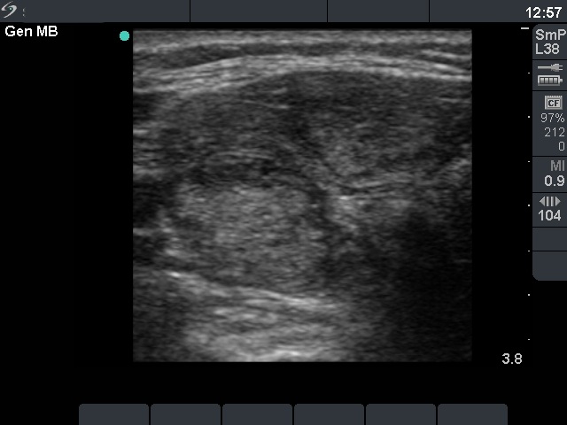
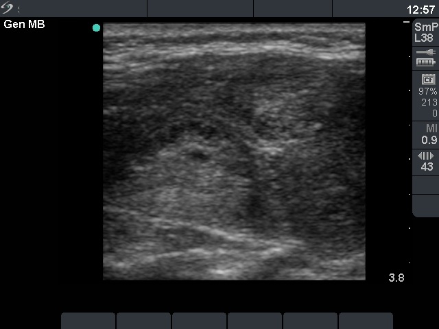
difference in the sonographic presentation of the two cases. There
are multiple hypoechogenic lesions in hypoechogenic background and
a solitary hypoechogenic nodule, Hashimoto's thyroiditis and oxyphilic
tumor, respectively.