|
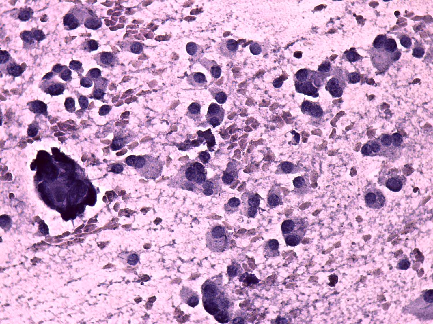
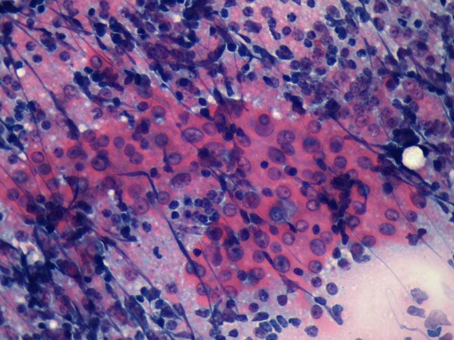
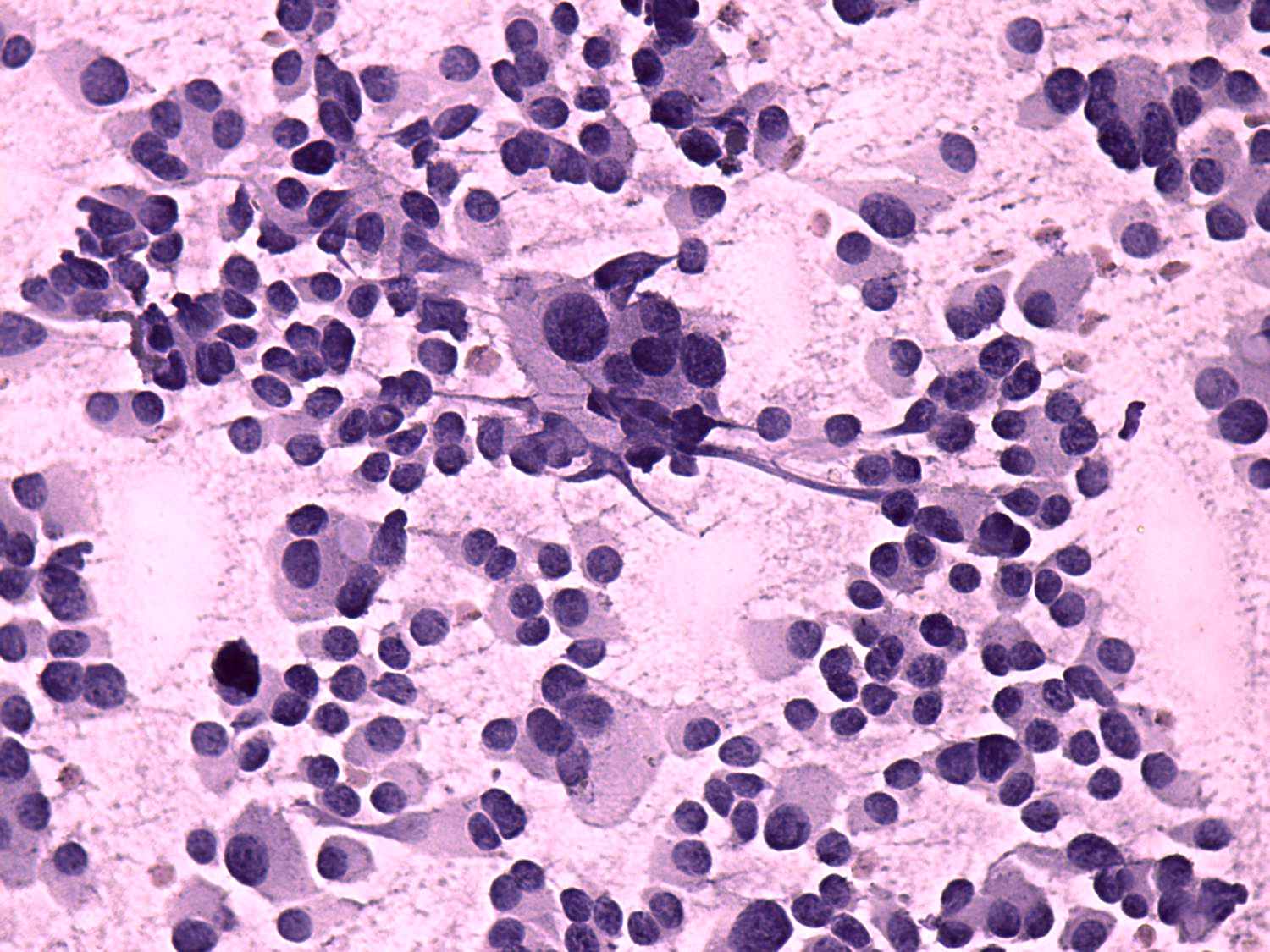
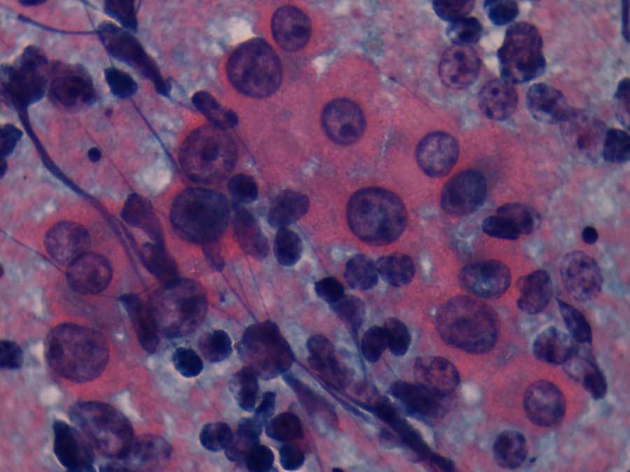
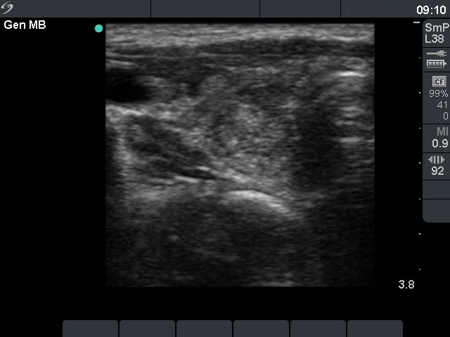
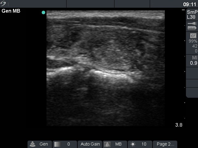
Compare the left
and right images. Histologically verified Hashimoto's thyroiditis is
demonstrated on the left, while a histologically proven coexisting
Hashimoto's thyroiditis and oxyphilic adenoma on the right one. In the
case of the tumor, single cells and microfollicles predominate the
smears, while in the case of thyroiditis follicular cells are found in
more cohesive structures. An even more important difference can be
observed analyzing the occurrence of lymphoid cells. As in this
example, no or only scattered number of lymphocytes are present on the
smear of a Hürthle-cell tumor. The extensive lymphocytic
infiltration practically excludes the possibility of a follicular type
Hürthle-cell tumor. In these cases, prominent nucleoli are found
completely opposite as in usual cases: oxyphilic cells contain large,
prominent nucleoli in this Hashimoto's case, while most oxyphilic cells
lack prominent nucleoli in this tumorous case. There is a small,
but important difference in the sonographic presentation of these two
cases. The lesion in question is larger in the case of oxyphilic tumor
than in the case of thyroiditis. This size, i.e. the maximal diameter
around 30 mm, practically excludes the possibility that this lesion
would be a secondary lobule. Naturally, the statement is conversely not
true: the small size of a lesion does not exclude the possibility of a
tumor. Moreover, in the case of oxyphilic tumor, the presence of a halo
sign and perinodular blood flow are strong arguments for the presence
of a follicular type tumor in such relatively large nodule. Taking all
of the above properties into account, the diagnosis of Hashimoto's
thyroiditis presented in left images was not difficult. In the right
case, the combination of cytological and sonographic data led to the
correct diagnosis.
|