Role of the teamwork - is there a coexistent oxyphilic tumor, too?
Chronic lymphocytic thyroiditis - Case 17
Chronic lymphocytic thyroiditis - Case 23: a concomitant oxyphilic adenoma


The presence of Hashimoto's thyroiditis is unequivocal in both cases. The question is whether a concomitant oxyphilic tumor is present or not. This chance is greater in the right case because of the presence of prominent nucleoli and condensed chromatin on the periphery of the nucleus. Nevertheless, there is no significant difference in the cytological patterns and it is hard to say that any of them would be definitively a Hürthle-cell tumor or definitively not.


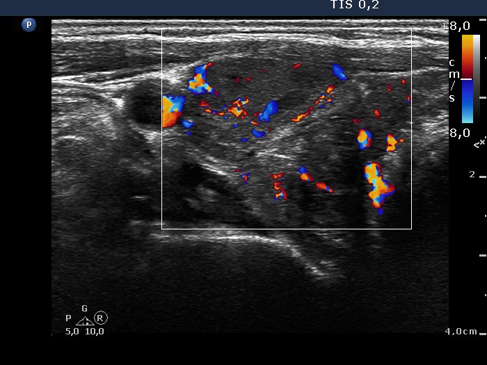
The thyroid is composed of several more hypoechogenic lesions. The ventral one was aspirated which presents halo sign and perilesional blood flow. On the other hand it is doubtful whether this area would be a nodule in a pathological sense. The multiplicity of discrete lesions argues against this possibility. Moreover, a follicular type oxyphilic tumor occurs generally in solitary nodules.

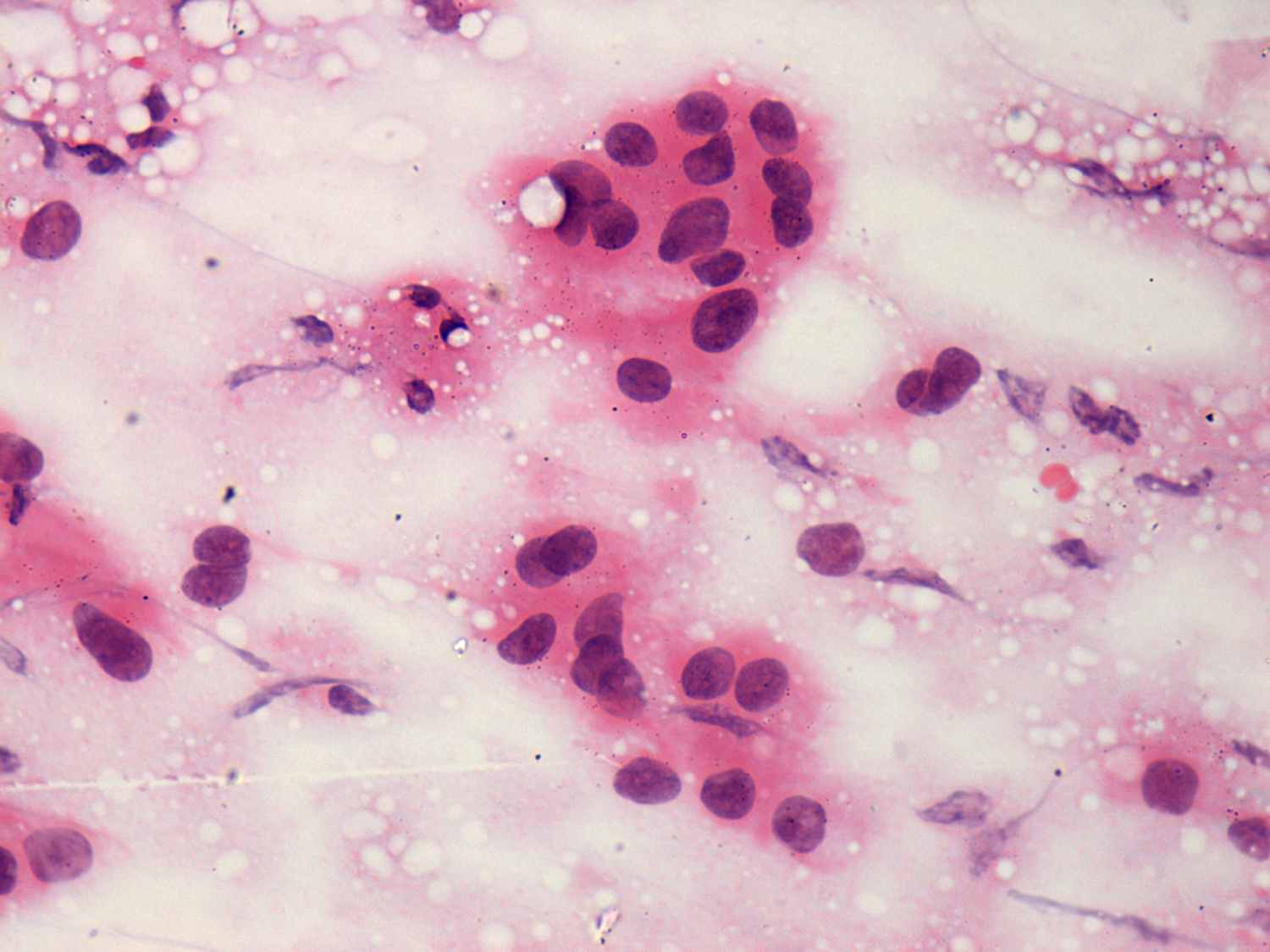
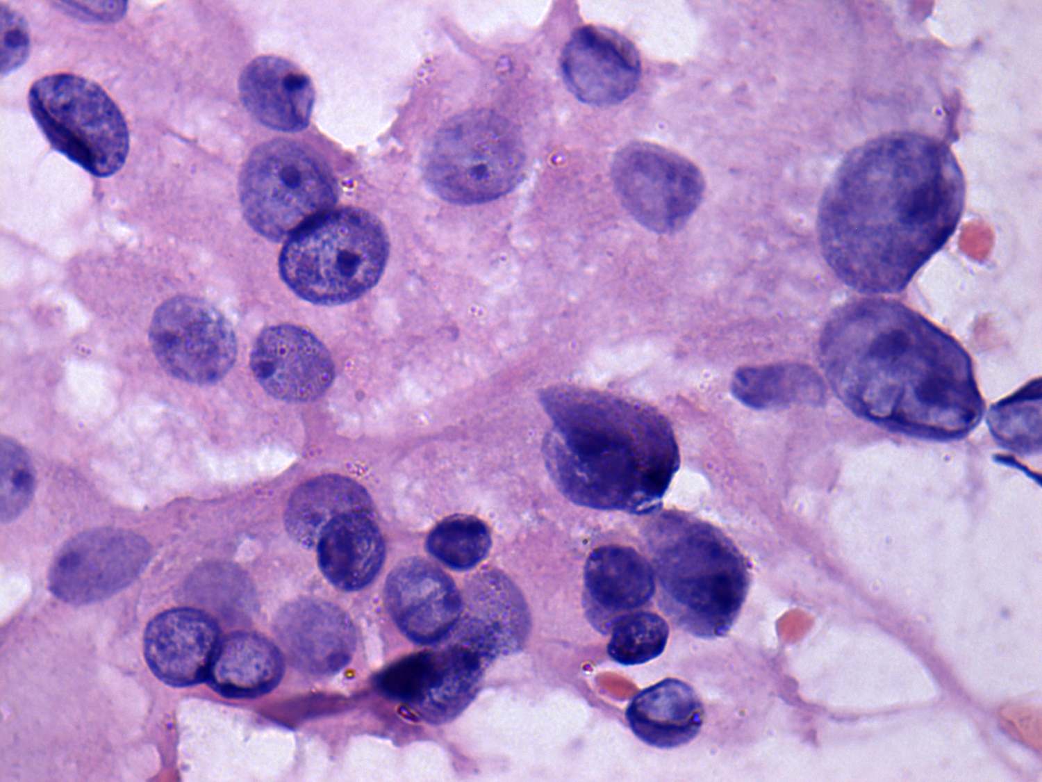
This is the typical presentation of a follicular type tumor. The lesion presented above is larger than a pseudolobule and is solitary.

Taking the sonographic and cytological pattern into account we gave the diagnosis of a Hashimoto's thyroiditis.

Taking the sonographic and cytological pattern into account we gave the diagnosis of a Hashimoto's thyroiditis and oxyphilic tumor.
mask