|
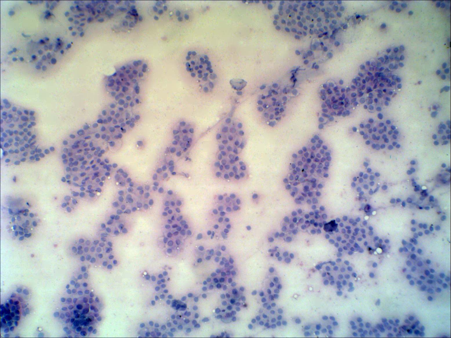
Two cell populations are present on the smear. We can find monolayered
sheets and normofollicles characteristic of Graves' disease.
The other pattern of thyrocytes is characterized by enlarged, elongated
cells presenting grooves. The chromatin structure of the latter cells
is not hyperchromatic. This pattern is suspicious for papillary cancer.
The lesion proved to be benign.
|
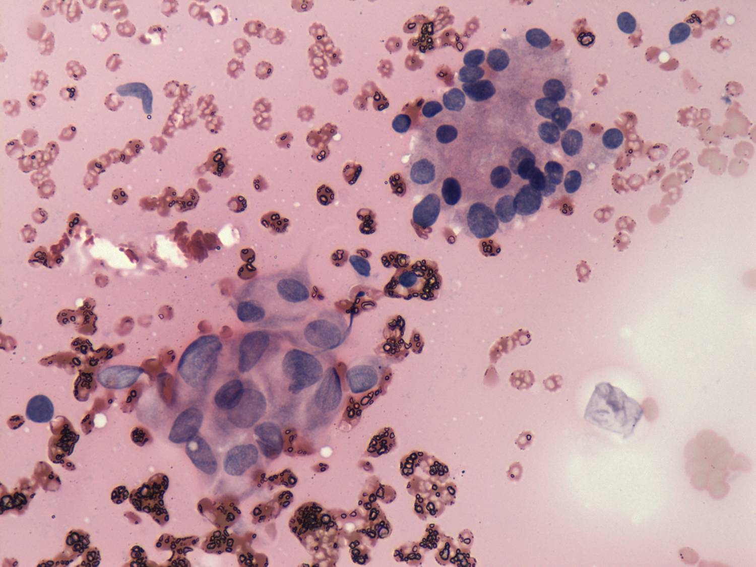
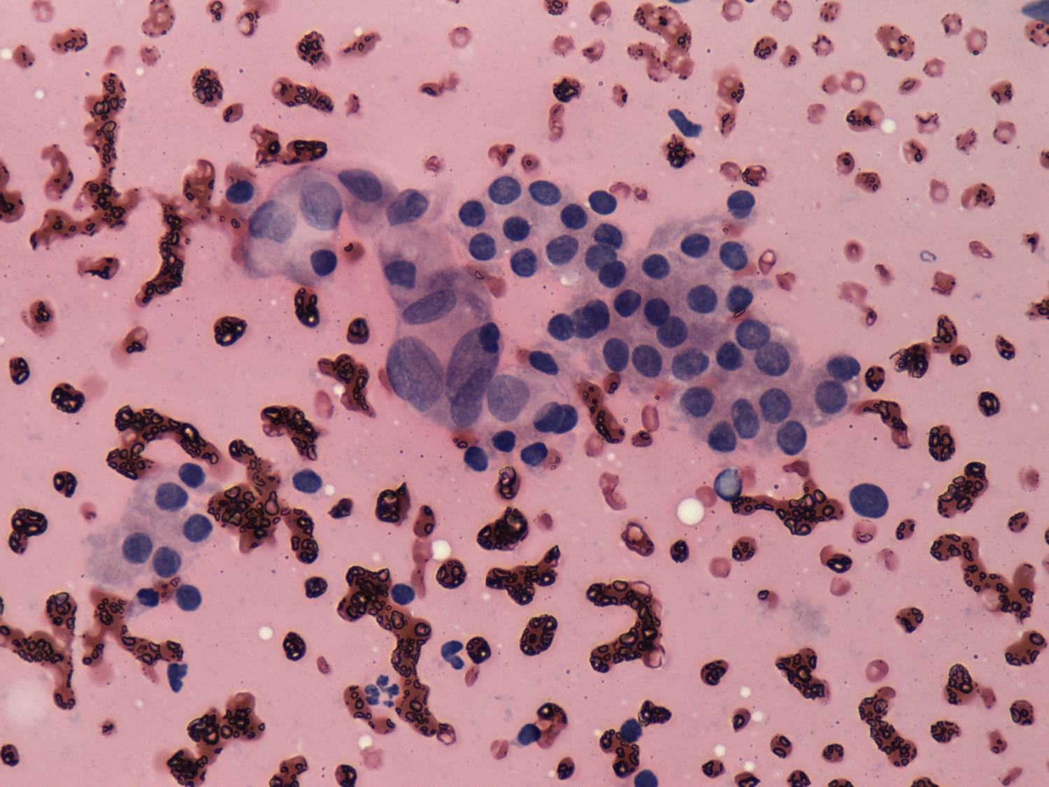
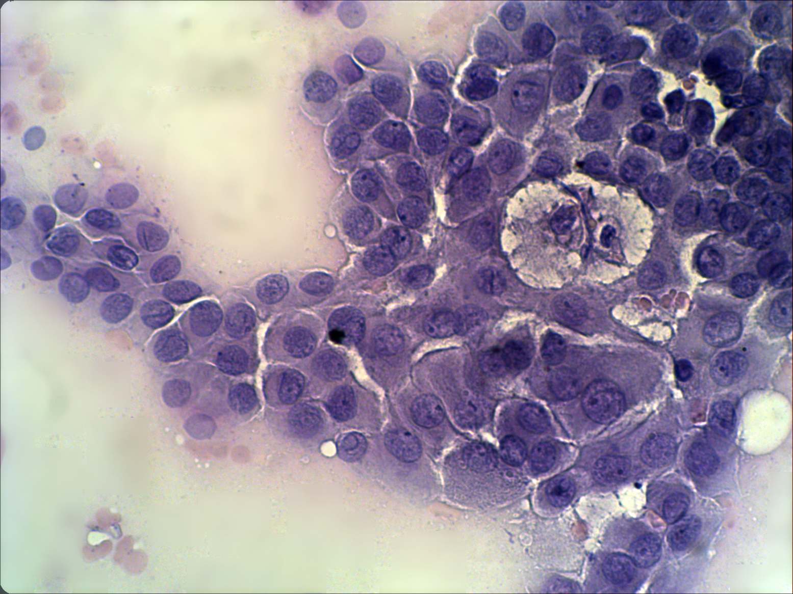
The following properties
raised the suspicion of papillary cancer. First, great proportion of
cells contained grooves. Second, follicular cells had abundant
cytoplasm and displayed distinct cellular borders. Nuclear crowding
could be observed. And finally, the presence of macrophages is an
unusual phenomen in Graves' disease.
Final histopathology disclosed papillary carcinoma.
|