
|
|
Papillary carcinoma - Case 53.
|
|


Clinical data: a 44-year-old man had been treated for Graves disease for 5 years. He requested a second opinion. Besides hormonal tests, a scintigraphy with normal findings was the only investigation performed at the beginning of the disease. He had no complaints.
Palpation: a moderately firm nodule in the left lobe was palpable.
Results of blood tests indicated euthyroidism on daily 10 mg methimazole (TSH-level 1.03 mIU/L, FT4 14.5 pM/L).
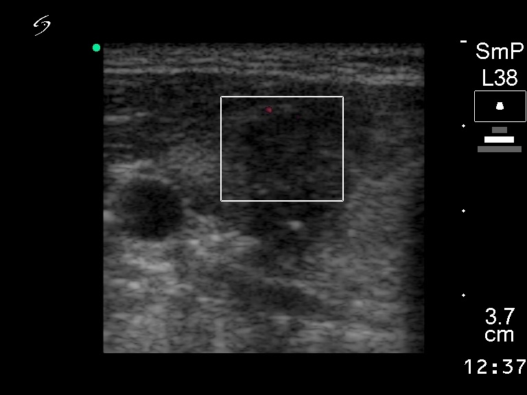
Ultrasonography revealed an echonormal thyroid. There was a hyperechogenic, inhomogeneous nodules with irregular borders in the right lobe. The vascularization was decreased within the nodule.
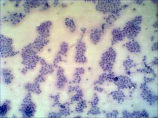
Cytological picture: no colloid can be found in the background. A very cellular picture. A great proportion of follicular cells exhibit inclusion or groove. Thyrocytes are arranged in clusters exhibiting nuclear crowding. There are many multinucleated giant cells on the smear.
Cytological diagnosis: papillary carcinoma.
Histopathology: papillary carcinoma.