
|
|
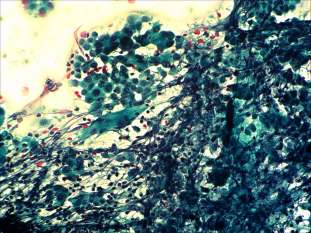
Papillary carcinoma - Case 54.
|
|


Clinical data: a 50-year-old woman was referred for an evaluation of a nodular goiter discovered on screening.
Palpation: the whole thyroid was moderately firm. A nodule was palpable in the upper pole of the left lobe.
Functional state: euthyroidism (TSH 1.73 mIU/L, FT4 17.0 pM/L, anti-TPO 742 IU/mL).
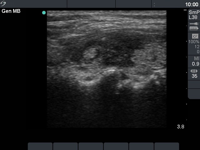
Ultrasonography: the thyroid was echonormal with hypoechogenic areas. The echogenicity index was around 25%.
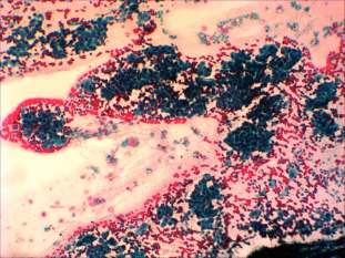
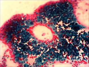
Cytological picture: no colloid in the background. Thyrocytes in loose clusters and dissociated. Thyrocytes exhibit oxyphilic metaplasia, prominent nucleoli, intranuclear inclusions and grooves. Note the multinucleated giant cells.
Cytological diagnosis: papillary carcinoma.
Histopathology: papillary carcinoma and Hashimoto's thyroiditis.
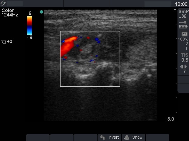
Comment: we gained multiple smears from the tumor. There were signs of Hashimoto's thyroiditis in the presented smear, too, while this was the only finding on other smears. The sonographic pattern of the tumor was identical with more active focus of Hashimoto's thyroiditis. The only difference was the size of this lesion which was larger than other lesions observed in the thyroid. In the case of Hashimoto's thyroiditis neither the irregularity of border, nor the increased vascularization has any relevance itself. The lesson to draw is the comparison of various lesions: if we detect a lesion which differs from others regarding the echogenicity, the size or the vascularization, it is advisable to perform FNAC.