Benign hyperplastic nodules - Case 1 |
 |
 |
|
Type 1 vascular pattern. It means neither perinodular nor intranodular
blood flow. This pattern decreases the likelyhood of a follciular
tumor.
|
| |
|
|
 |
 |
|
Type 2 vascular pattern. It means perinodular blood flow. This is the
typical presentation of a follicular tumor. The presence of perinodular
blood flow in a solitary nodule larger than 20 mm greatly increases the
likelyhood of follicular tumor.
Conversely, the lack of halo sign and perinodular blood flow almost
excludes the possibility of follicular tumor.
|
| |
|
Benign hyperplastic nodules - Case 17 |
 |
 |
|
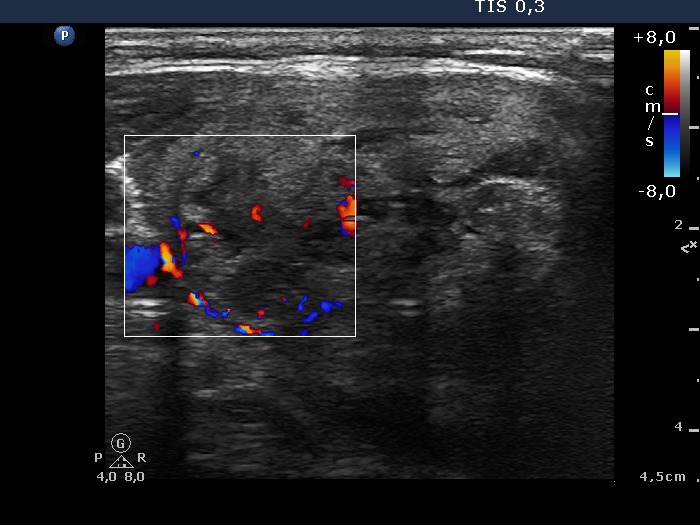
Type 3 vascular pattern. It means intranodular blood flow. Although
such pattern significantly increases the risk of papillary cancer, this
sign has itself has very limited practical value in diagnosing
papillary carcinoma in contrast with the presence of microcalcification
or blurred borders.
|
| |
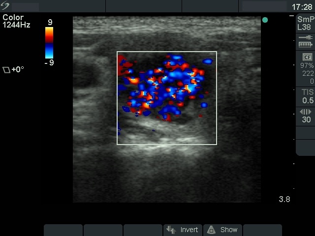
Benign hyperplastic nodules - Case 5 |
 |
 |
|
Combined type 2 and type 3 vascular pattern, i.e. the nodule presents
both perinodular and intranodular blood flow. This type of
vascularization is frequently observed in autonomously functioning
adenoma.
|
| |
|
Benign hyperplastic nodules - Case 8 |
|
|
|
| Incomplete
type 2 and type 3 vascular pattern, i.e. the nodule presents an
incomplete perinodular and displays an intranodular blood flow. In gray
scale mode, the presence of halo sign is doubtful. The perinodular
blood flow and the halo are sonographic signs of a capsule. We
frequently find these sings incompletely, i.e. not all around a nodule.
Is such cases we can neither disclose nor exclude the possibility of a
follicular type tumor. |
| |
|
Benign hyperplastic nodules - Case 30 |
 |
 |
|
This case illustrates what really "blood flow" means. By Doppler mode
we measure and visualize the flow of fluid irrespective of its nature.
In this case the flow of cystic fluid and not that of blood is
presented.
|


