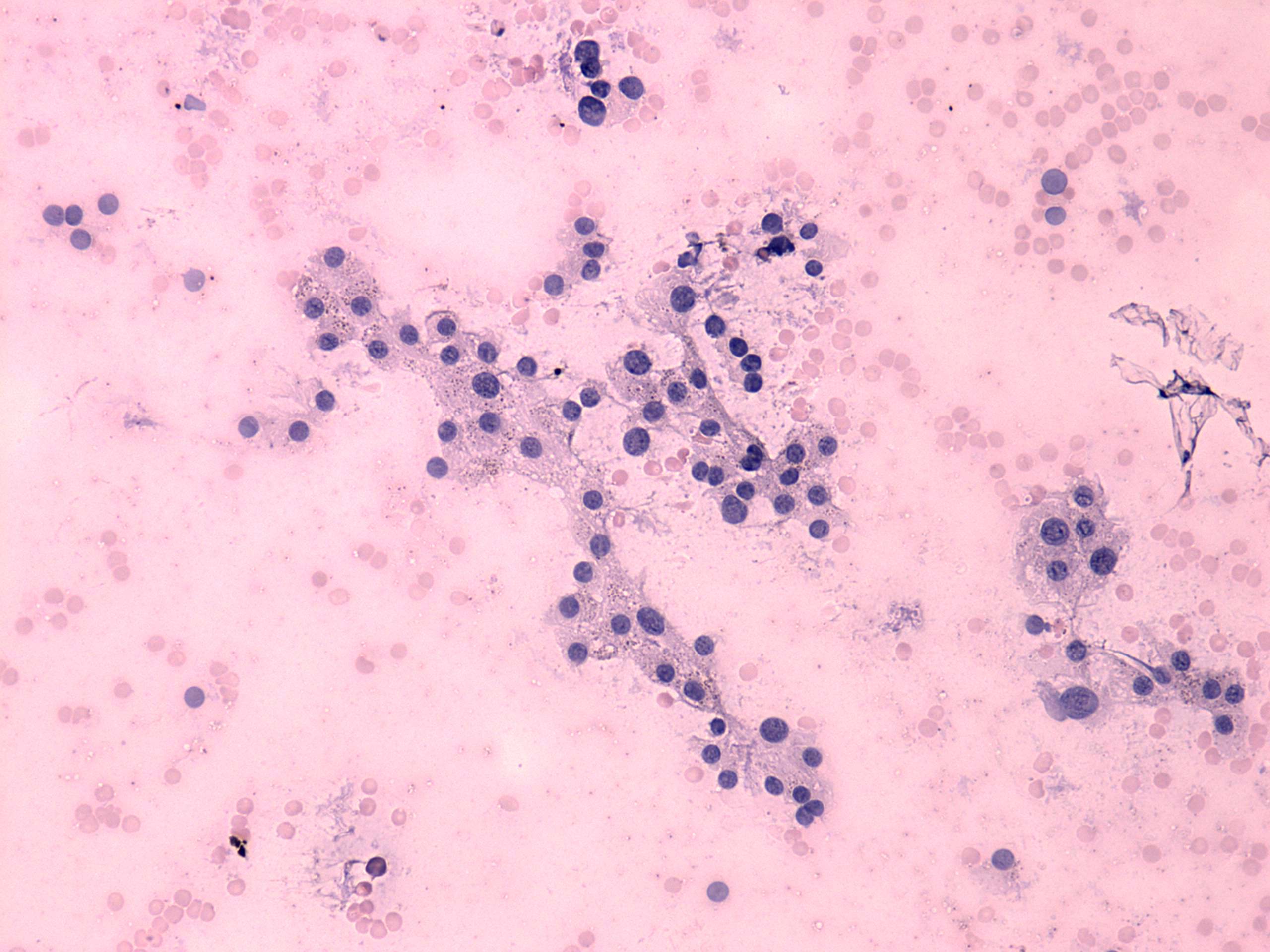
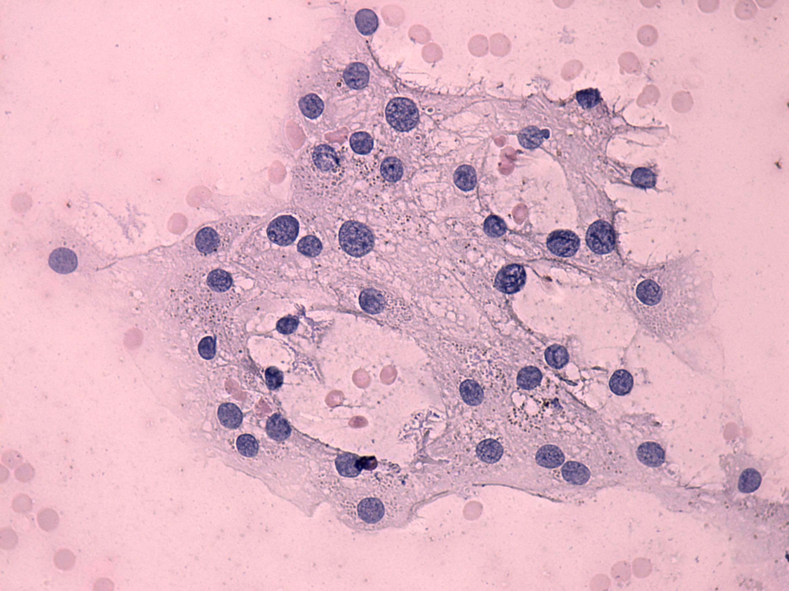
The cytological pattern presents signs of hyperfunction: anisonucleosis
without plemorphism and vacuolization.
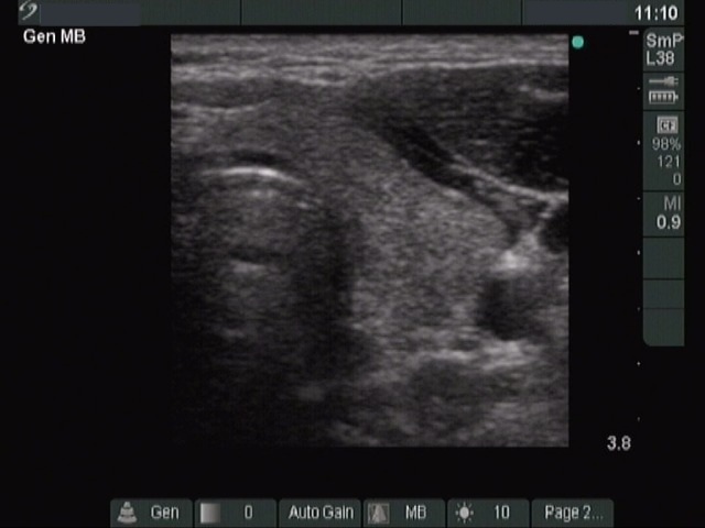
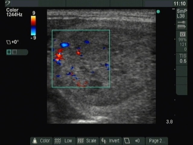
Right lobe
Left lobe
Typical sonographic presentation of an
autonomously functioning nodule: the lesion displays a halo sign and
combined type 2 and 3 vascular pattern, while the contralateral lobe is
atrophic. The latter is caused by the low TSH level.
The patient was already euthyroid, but her TSH was relatively low (0.51
mIU/L). Taking the sonographic and cytologic pattern into account, the
probability of an autonomously functioning nodule was very high which
was proven by scintigraphy.
Naturally, the title of this table is only a joke. The
only way to diagnose an autonomously functioning nodule is to perform
isotope examination.