|
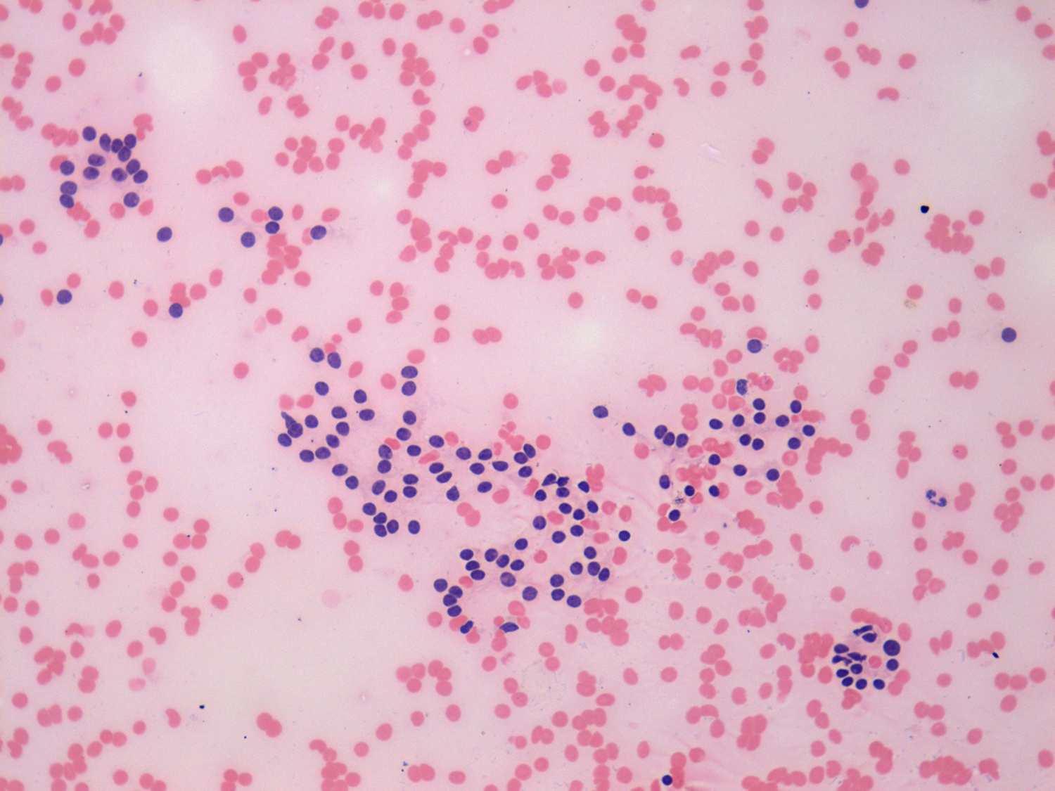
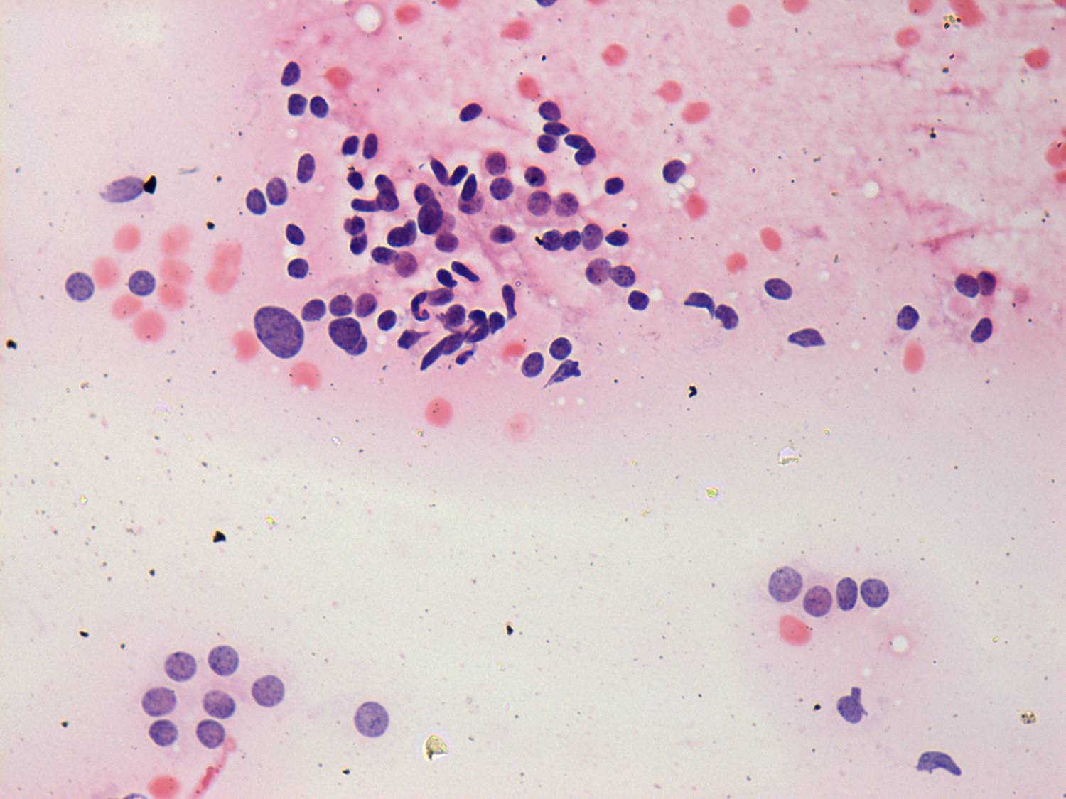
This is not a simple microfollicular pattern, because normofollicles
predominate the smear. Moreover follicular cells do not contain
prominent nucleoli. Nevertheless, the cytological picture is suspicious
for follicular tumor.
|
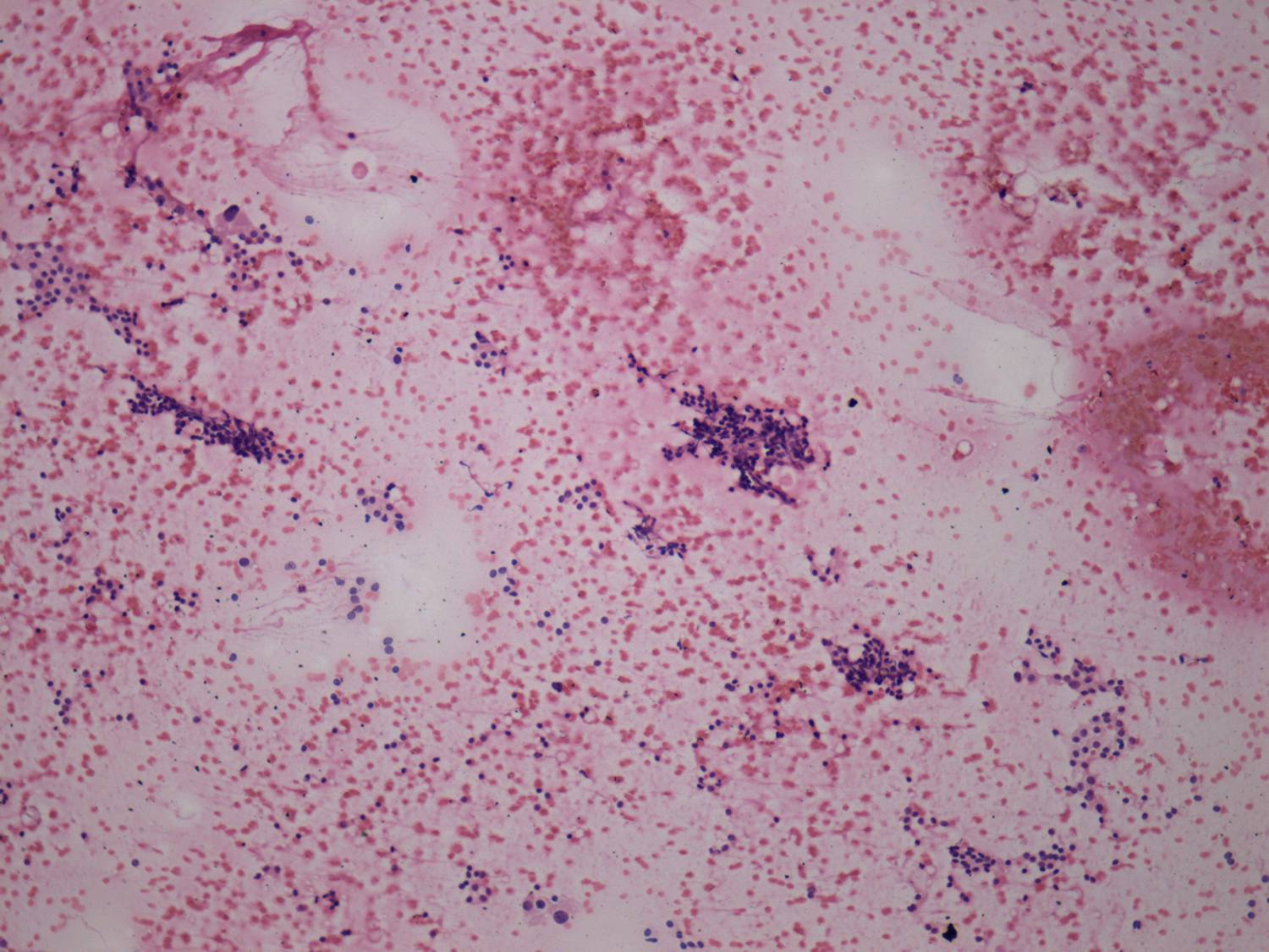
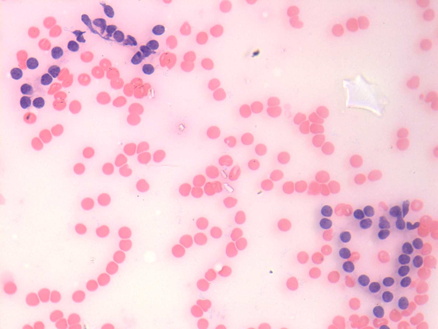
This is also not a classical microfollicular pattern. Thyrocytes are
partly found in hyperplastic papillary fragments. Nevertheless, first
of all in low power field the pattern raises the possibility of
follicular tumor.
|
|
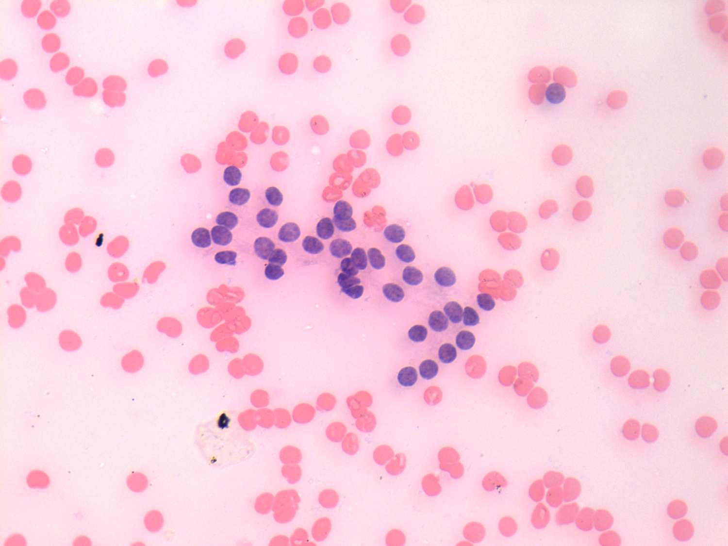
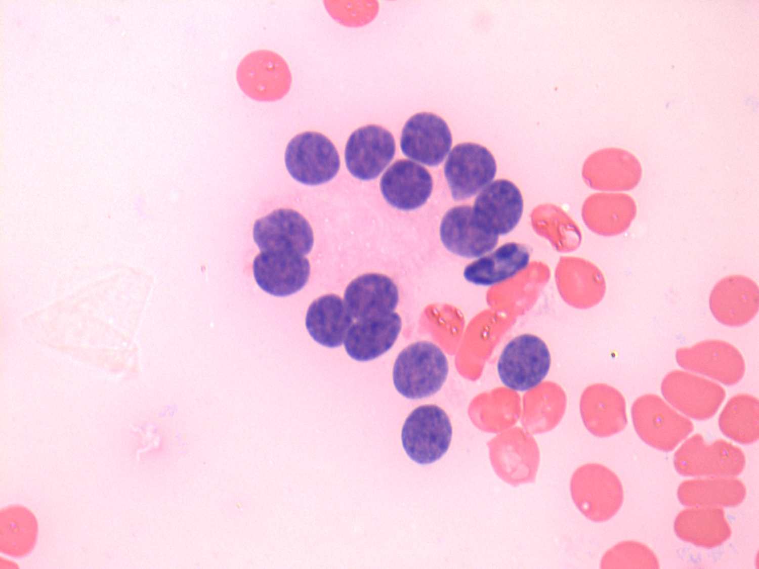
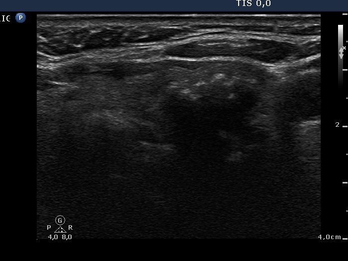
Taking the ultrasound pattern into account, we could avoid a false
diagnosis of follicular tumor, our combined cytological-sonographic
diagnosis was benign follicular proliferation. Final histopathology
disclosed benign hyperplastic nodule.
|
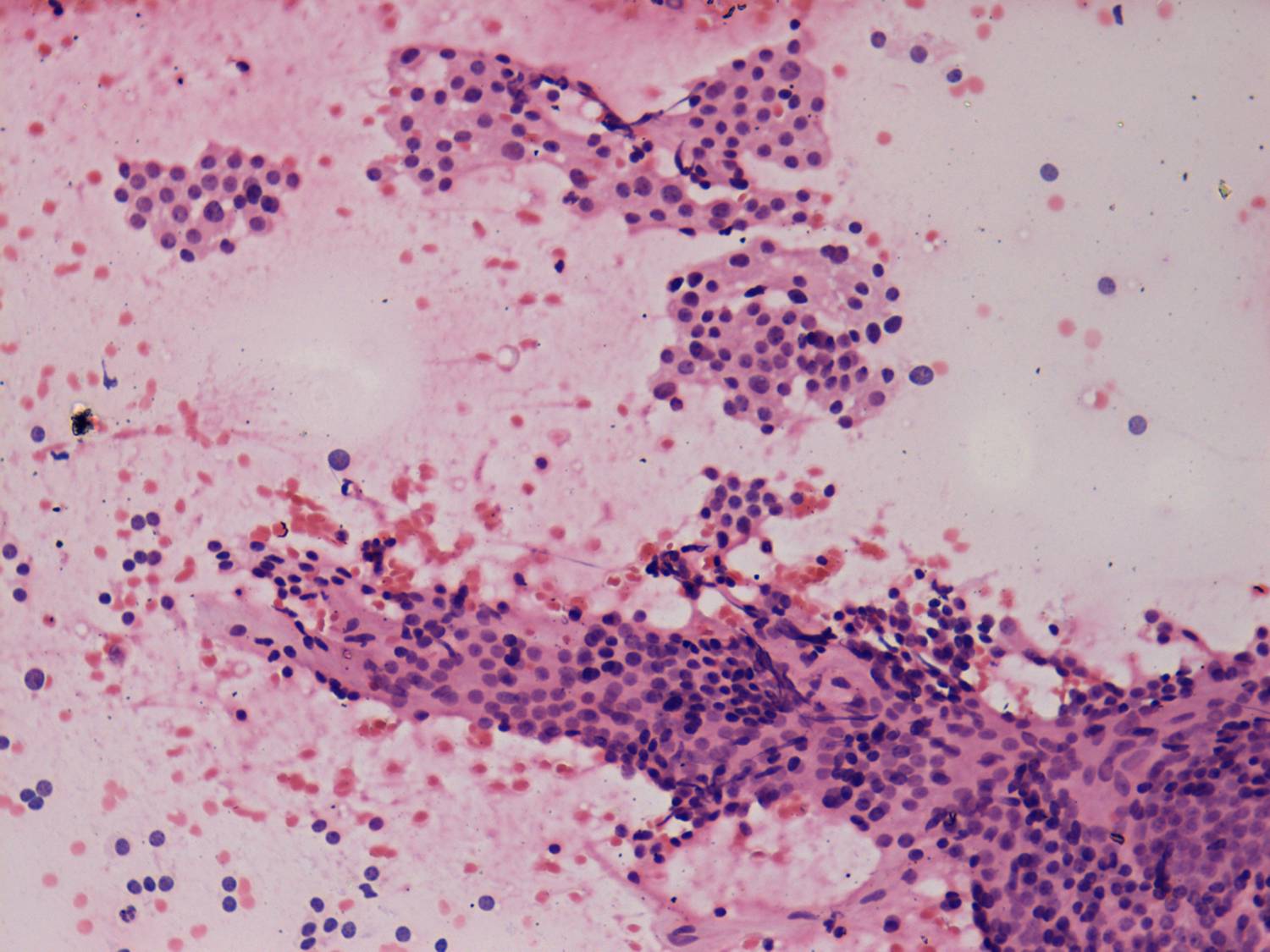
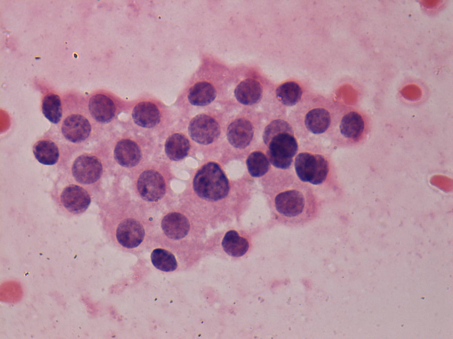
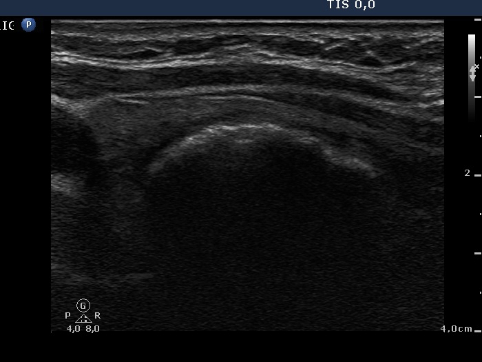
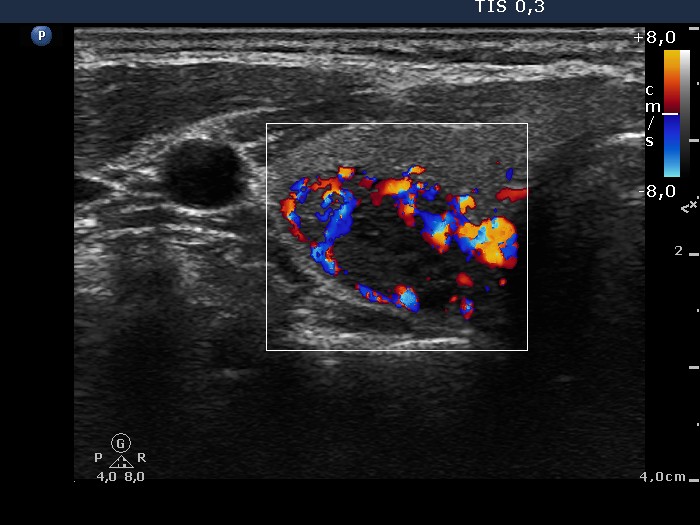
Taking the sonographic and the cytological pattern into
account our diagnosis was follicular tumor. |