Benign hyperplastic nodule - Case 2 |
|
 |
 |
 |
 |
 |
 |
 |
 |
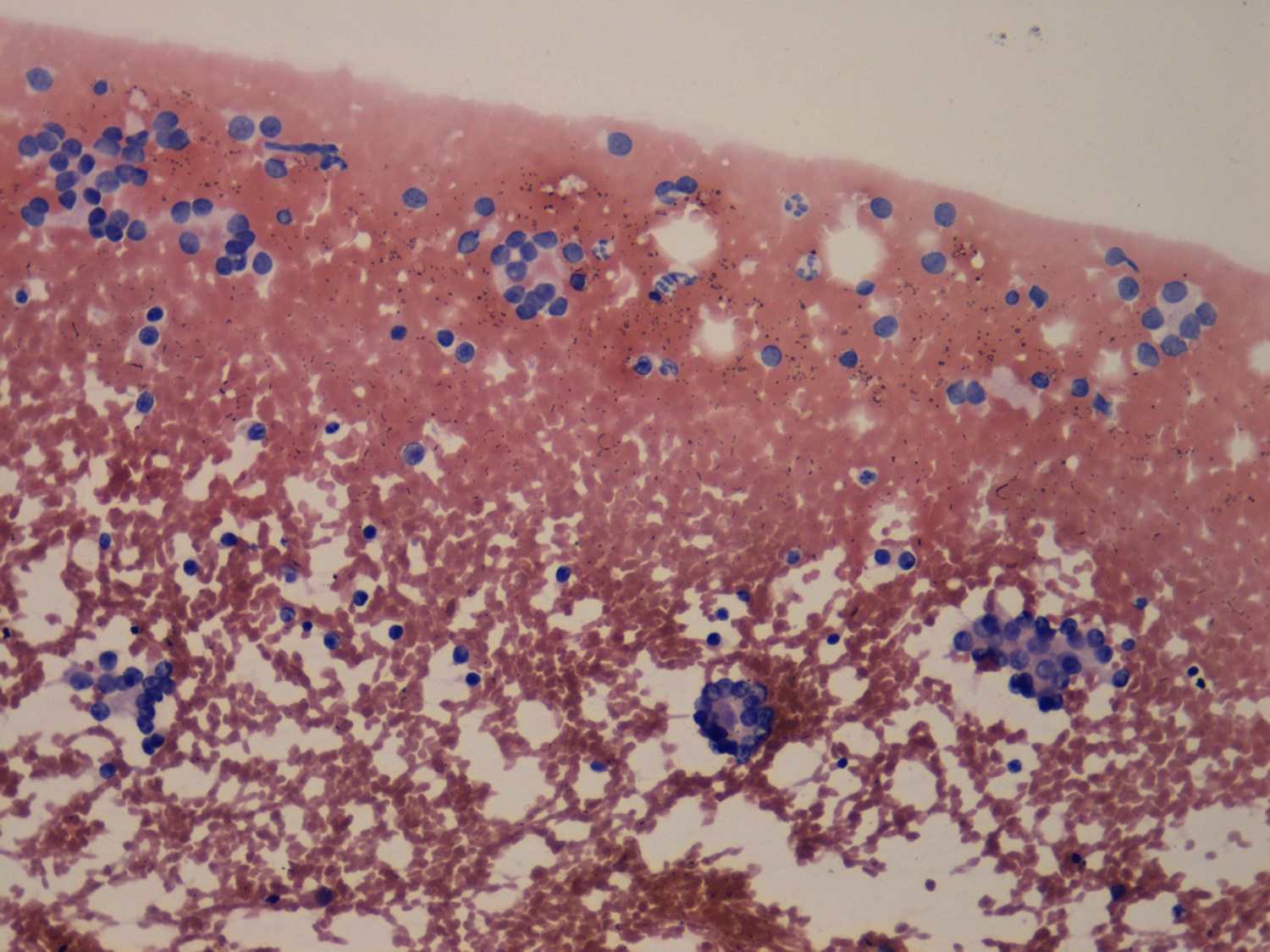
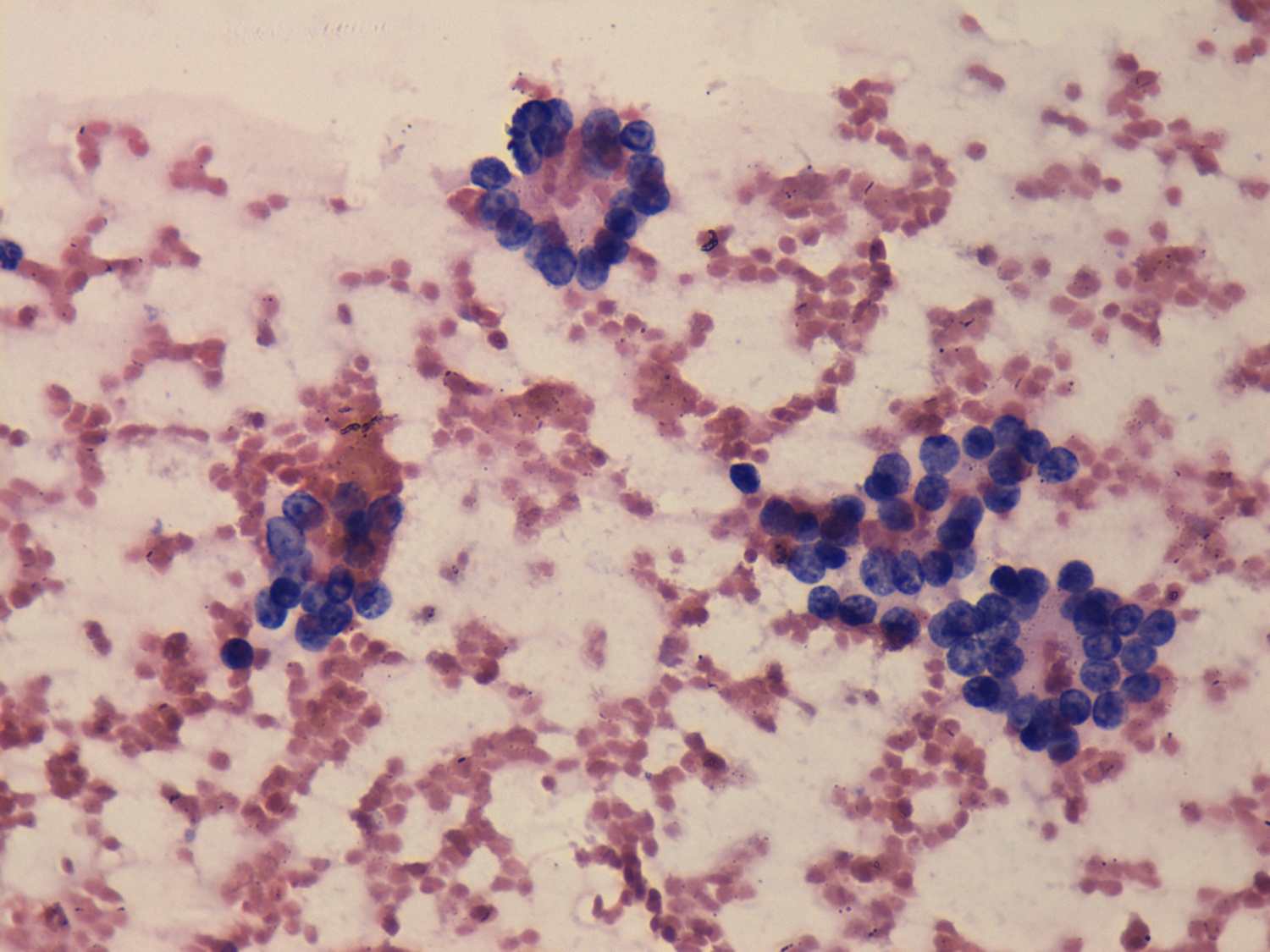
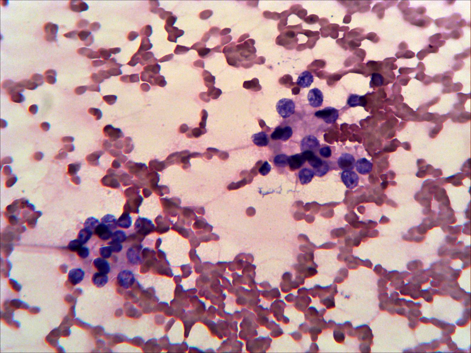
The pattern is identical to that seen in follicular tumor: there is
no colloid in the background, microfollicles predominate the smear, and
thyrocytes present prominent nucleoli.
|
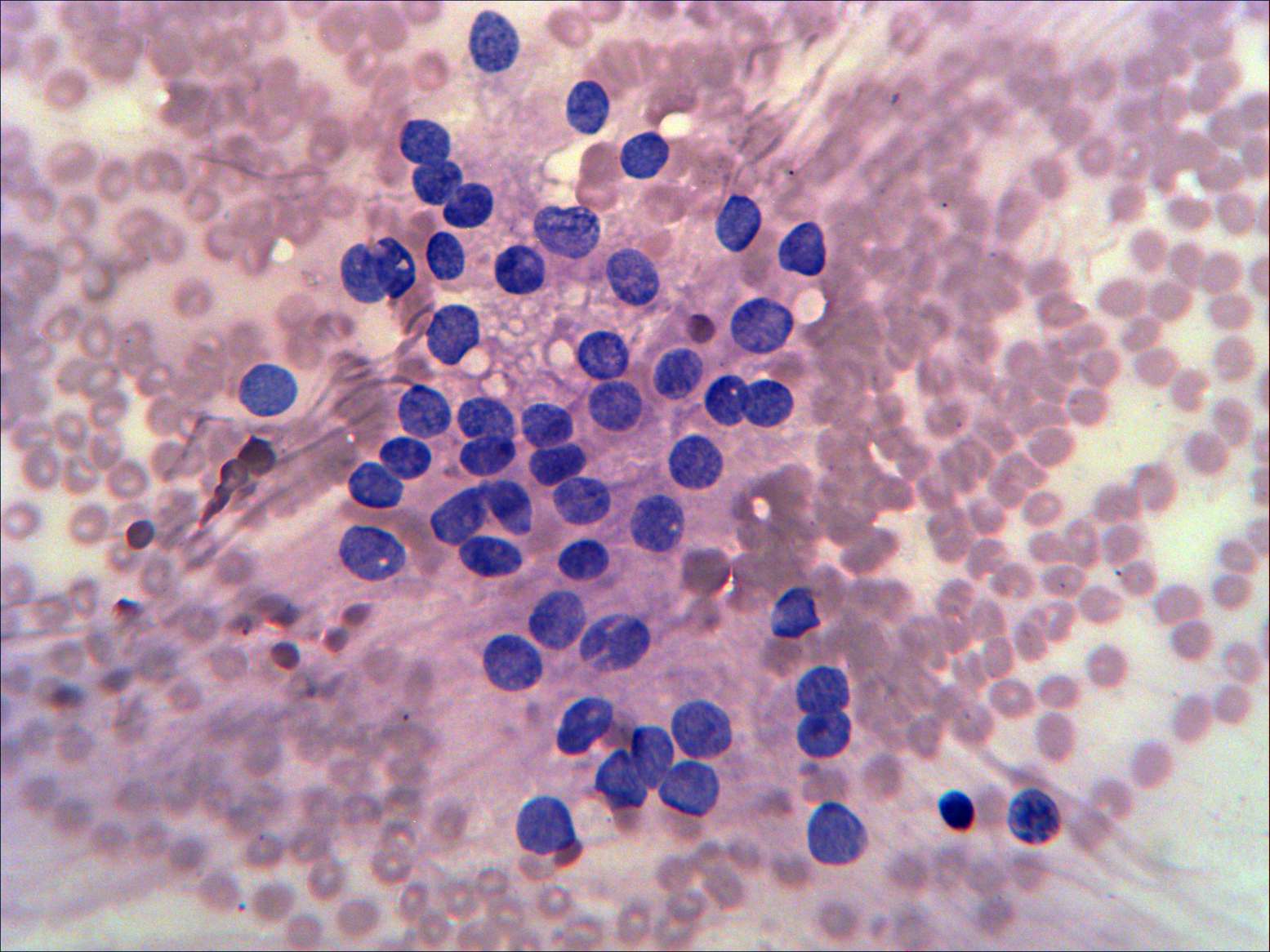
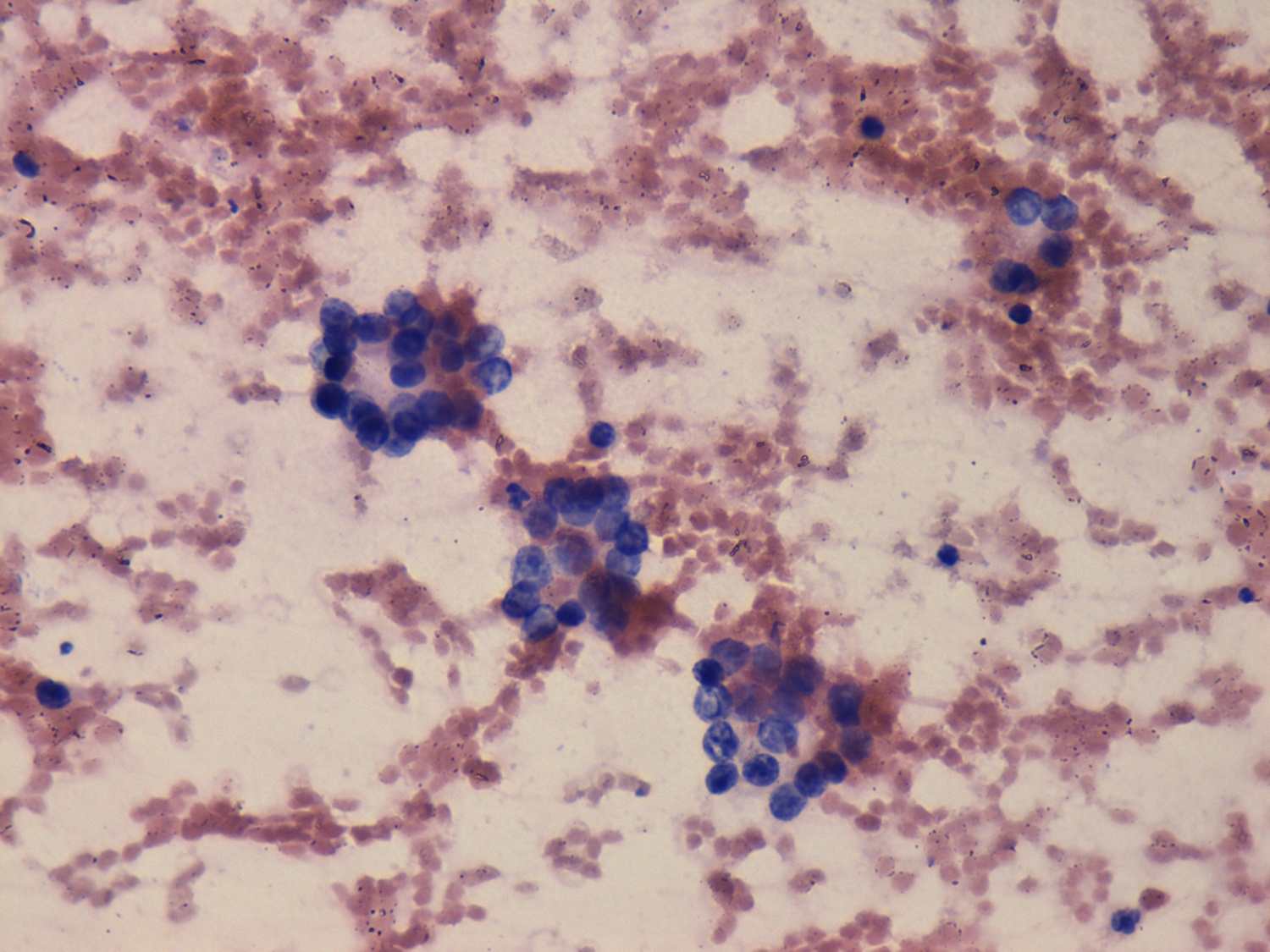
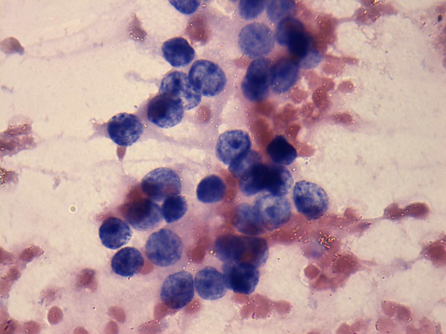
Micro- and normofollicles predominate the smear. Follicular cells have
coarsely granular chromatin structure. This cytological pattern also
corresponds to a follicular tumor.
|
 |
 |
 |
 |
|
This ultrasound pattern significantly decreases the possibility of
follicular tumor. There is no halo sign and the lesion displays a
type 1 vascular pattern; it means there were no sonographic signs of a
capsule which is an essential part of follicular tumor.
|
The presence of a halo is more than doubtful. On the other hand, a type
2
vascular pattern is a very strong argument for the presence of a
capsule.
This pattern in a solitary nodule is almost diagnostic for follicular
tumor. |
|
Taking the ultrasound pattern into account, we could avoid a false
diagnosis of follicular tumor, our combined cytological-sonographic
diagnosis was benign follicular proliferation. The patient was operated
because she harbored another large nodule. Final histopathology
disclosed benign hyperplastic nodule.
|
Our diagnosis was follicular tumor. Final histopathology disclosed
minimally invasive follicular carcinoma.
|

