Role of the teamwork in diagnosing papillary carcinoma (VI)
Papillary cancers - Case 65

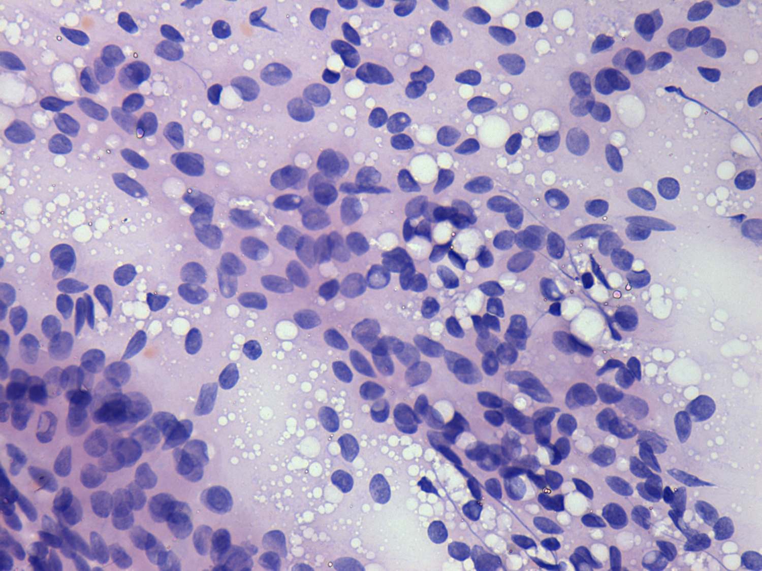
On low power field we had little if any doubt that this is a colloid goiter. On the other hand we have found two suspicious signs on high power. First, a few inclusions were found on the smear. (The intranuclear hole marked with red corresponds to a suspicious inclusion while the remaining holes do not. For explanation see case history). Second, there were several cell groups with a disarranged structure, wiht nuclear crowding and overlapping.

Ultrasonography


This pattern is reassuring: an echonormal nodule with halo sign and perinodular blood flow. It must be a follicular tumor with all probability.


We gave a common cytological-sonographic diagnosis: follicular tumor with less than the average risk for malignancy. Final histopathology disclosed follicular variant of papillary cancer.
This case illustrates that the combination of sonographic and cytological data may also lead to false diagnosis. Nevertheless, in most cases the combination of these tools has cumulative effect.

mask