
|
|
Papillary carcinoma - Case 65.
|
|


Clinical data: a 36-year-old woman referred for evaluation of a nodule discovered for 4 months.
Palpation: a firm nodule in the left lobe.
Functional state: euthyroidism with TSH-level 1.66 mIU/L, FT4 13.1 pM/L.
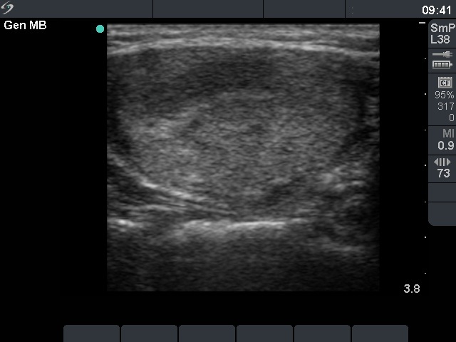
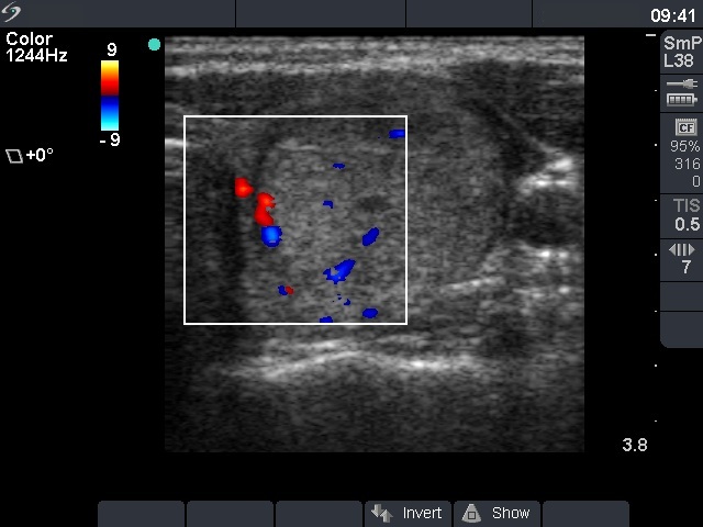
Ultrasonography: an echonormal nodule in the left lobe with halo sign and perinodular blood flow.
Combined ultrasonographic-cytological report: follicular tumor with less than the average risk for malignancy.
Histopathology: follicular variant of papillary cancer according to the nodule in the left lobe with metastasis to the right lobe.
Comments.
-
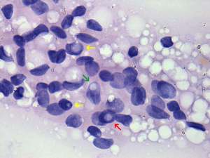
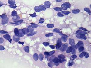
The nodule as a whole is proved to be papillary cancer on histopathology and almost the entire lobe was consisted of the nodule. It means that FNAC was falsely negative, not because of error of gaining adequate material but because of the error of the interpretation.
Nevertheless, it is very hard to reconsider our original diagnosis, but we have to do it. The presence of colloid, that of pycnotic nuclei and the cellular pattern as a whole argue for a benign lesion. There are two possible signs of papillary cancer on the smear: firstly, the presence of a few inclusions and secondly, the irregularity of several clusters. Both of these properties are frequently found in benign colloid goiter. The clue is the occurrence of inclusions in greater than the acceptable proportion. -
Papillary cancer is found only very rarely in echonormal nodules.
.