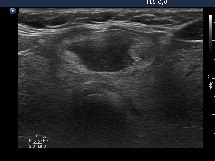
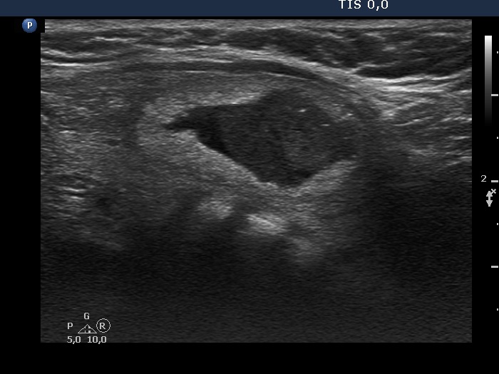
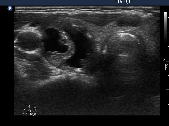
Benign cystic-colloid goiter (cytological diagnosis) |
 |
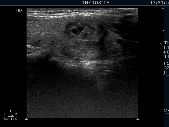 |
There are several pale granules and lines in the solid part of the nodule (left image), while the right image demonstrates the presence of posterior back wall enhancement figure in the dorsal wall of the small cystic areas.
|
| |
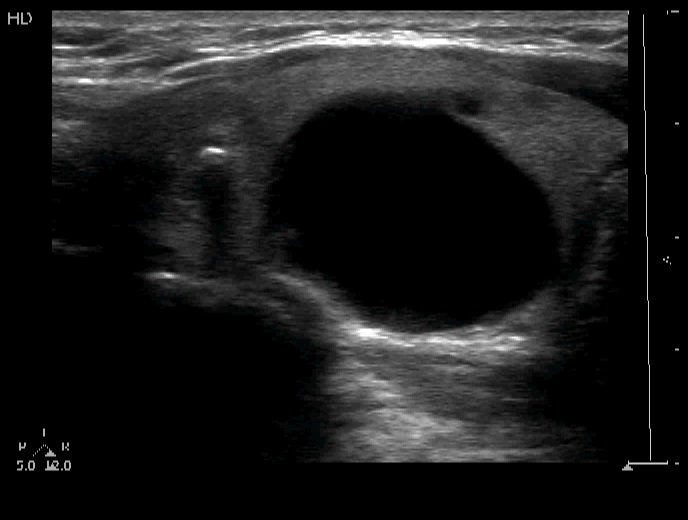
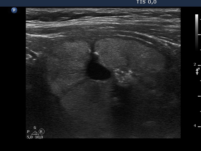
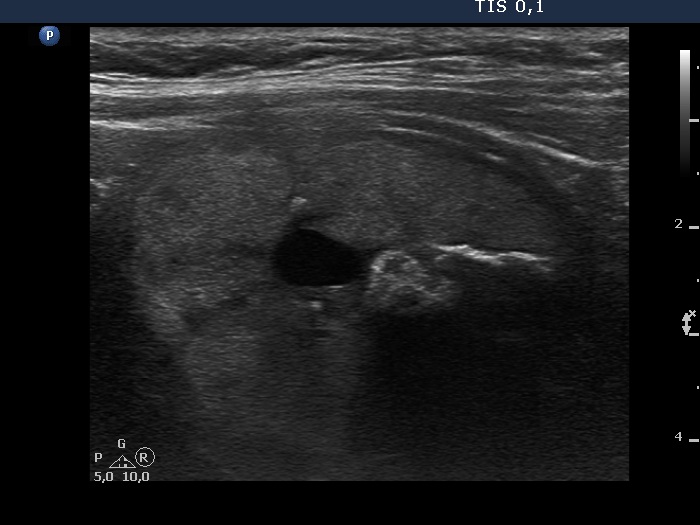
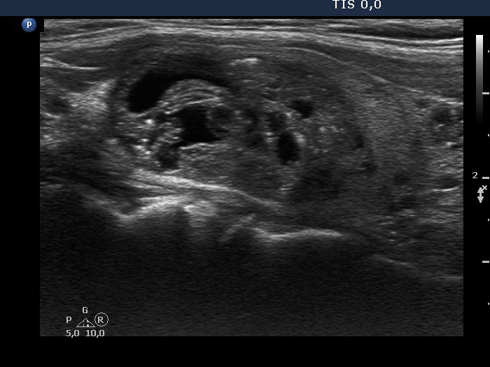
Follicular adenoma (histological diagnosis) |
Before aspiration |
 |
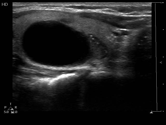 |
After aspiration 6.5 ml cystic fluid
|
 |
 |
The granules in the solid part lower to the cystic area are punctate echogenic foci (microcalcifications).
|
| |
|
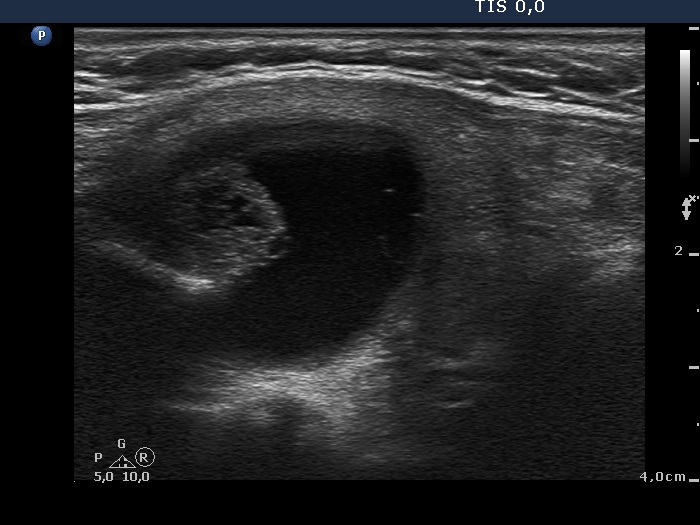
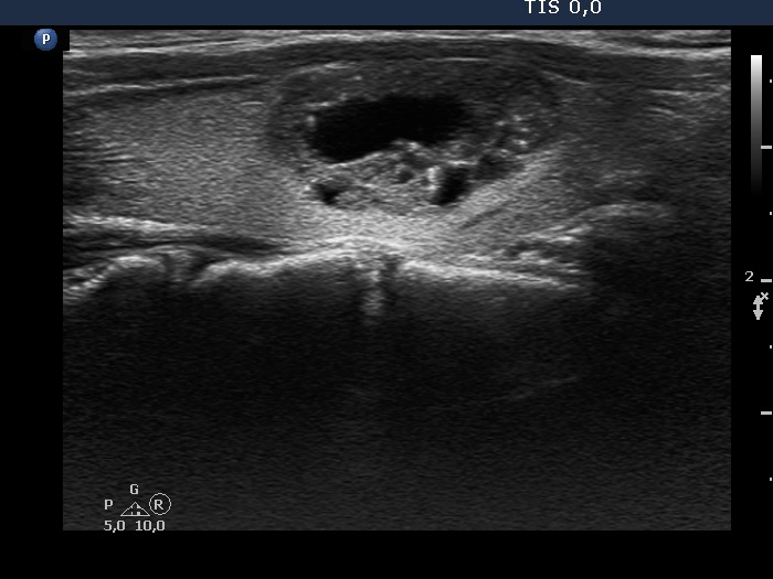
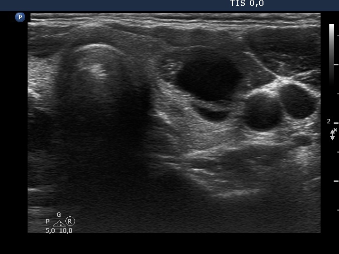
Benign cystic-colloid goiter (cytological diagnosis) - case 1473
|
 |
 |
 |
 |
The synchronous presence of bright granules and lines are ultrasound presentations of the connective tissue.
|
| |
|
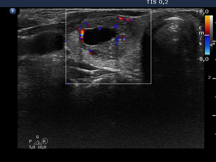
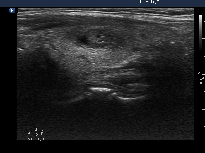
Benign cystic degeneration (cytological diagnosis) - case 1669 |
 |
 |
 |
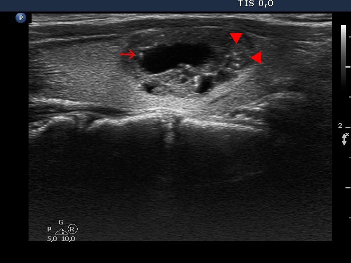 |
The lesion contains various figures: there is a comet-tail artifact marked with arrow, there are optical artifacts caused by posterior enhancement in the back wall of the cysts. However, the categorization of hyperechogenic granules (arrowheads) in the solid part of the lesion is obscure; these might be punctate echogenic foci (microcalcifications).
|
| |
Papillary carcinoma (histological diagnosis) - case 853 |
 |
 |
The nodule has several bright and pale granules without a tail and without the synchronous presence of hyperechogenic lines; therefore we have to conclude that these figures are punctate echogenic foci.
|
| |
Benign cystic degeneration (cytological diagnosis) - case 808 |
Before aspiration |
 |
 |
After aspiration of 5 mL cystic fluid
|
 |
 |
The bright granule within the solid part has a short tail; therefore it is a colloid crystal with great probability.
|
| |
Benign colloid goiter (cytological diagnosis) |
 |
 |
The echonormal large nodule presents three different coarsely calcified foci: a small one close to the ventral wall (in the left image), an amorphous which contains tiny granules in the central part (in both images), and a linear in the lower part of the lesion (in the right image).
|
| |
Benign cystic-colloid goiter (cytological diagnosis) |
 |
 |
Besides various forms caused by posterior acoustic enhancement and a few unequivocal colloid crystals, there are non-specific pale granules and lines and several bright large amorphous figures in the ventral solid part of the nodule.
|
| |
|
Benign cystic-colloid goiter (cytological diagnosis)
|
 |
 |
The hyperechogenic granules within the ventral solid part might be microcalcifications. The echogenic figures in the solid area cannot be the consequence of posterior enhancement because they are in front of the cystic area. Note that the nodule has in fact a moderately hypoechogenic solid part which seems to be echonormal dorsal to the cystic fluid because of the acoustic enhancement dorsal to the fluid.
|
| |
|
