
Benign nodular hyperplasia - Case 46. (ultrasonographic picture 2)
|
|

|
|
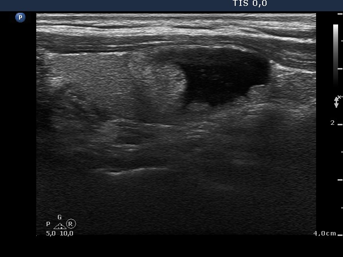
Right lobe, longitudinal scan. There is an echonormal solid part in the upper pole (left in the image) and several smaller ones in the dorsal part of the nodule.