
|
|
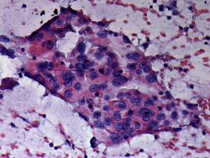
Chronic lymphocytic thyroiditis - Case 39.
|
|


Clinical data: a 48-year-old woman with hypothyroidism replaced with 50 microgram levothyroxine was sent for aspiration cytology because of a suspicious nodule found on ultrasound.
Palpation:
no abnormality.
Functional state: euthyroidism on daily 87.5 microgram levothyroxine therapy (TSH-level 0.39 mIU/L).
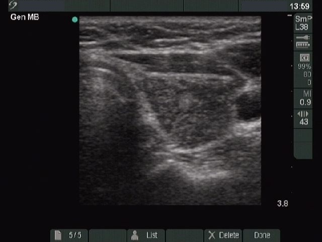
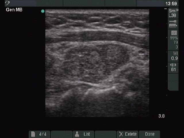
Ultrasonography. The two lobes differed in echo pattern. The right lobe (upper image) was echonormal and contained small hypoechogenic foci with an echogenicity index of less than 5% while the left lobe (lower images) was composed of a central hypoechogenic area surrounded with an echonormal rim, therefore it resembled a large hypoechogenic nodule comprising great proportion of the lobe . On the other hand the lesion had irregular borders and showed a heterogeneous pattern and similar smaller areas were found elsewhere in the lobe.
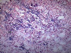
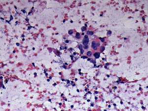
Cytological diagnosis: benign Hashimoto's thyroiditis.
Comment. The ultrasound pattern is remarkable, the left lobe highly resembles a pattern of a large hypoechogenic nodule. On thorough analysis of the images and the video, it seemed more likely that this area is not a nodule in a pathological sense but only more active focus of the underlying thyroiditis. The differentiation between a nodule in pathological sense and a hypoechogenic focus of Hashimoto's thyroiditis is not always possible, even in this case it not fully unequivocal.