
Papillary carcinoma - Case 39.
First investigation (ultrasonographic picture 3)
|
|

|
|
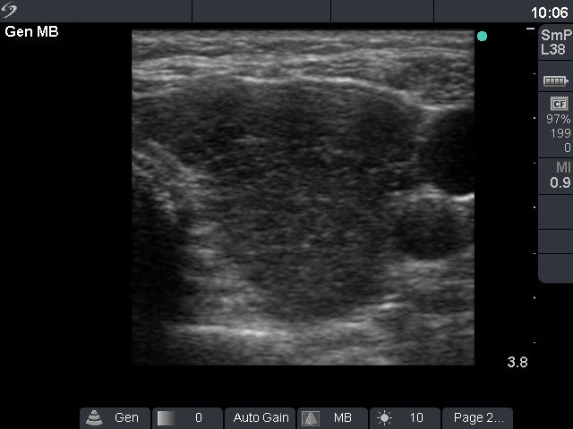
Left lobe, horizontal scan. The pattern is similar in the left lobe. There is no circumscribed lesion within the lobe.