Benign nodular hyperplasia - Case 20. (ultrasonographic picture 2)
|
|

|
|
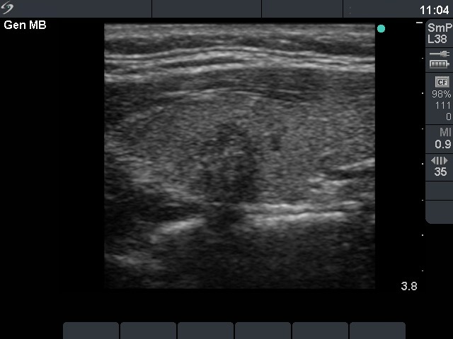
Left lobe, longitudinal scan. The nodule contains microcalcifications.

|
|
|
|
Left lobe, longitudinal scan. The nodule contains microcalcifications.