
|
|
Benign nodular hyperplasia - Case 34.
|
|


Clinical presentation: a 51-year-old woman with a thyroid nodule discovered by her family physician on a routine examination.
Palpation: a nodule in the right thyroid.
Functional state: euthyroidism (TSH 1.07 mIU/L).
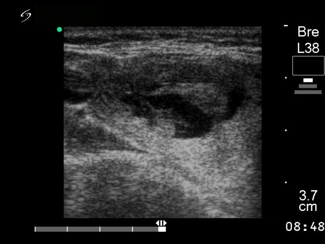
Ultrasonography:: a mixed hypoechogenic-echonormal nodule in the right lobe. The nodule presented neither a halo sign on the gray scale nor perinodular blood flow on the Doppler mode.
Combined cytological-sonographic diagnosis: a benign colloid goiter.
The patient wished to be operated and surgery was performed.
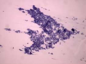
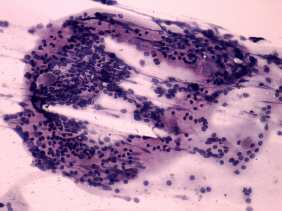
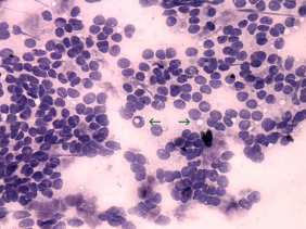
Histopathology: benign hyperplastic nodule. Chronic lymphocytic thyroiditis.
Comment: the cellular picture with microfollicular proliferation is very impressive. There are three properties standing against the possibility of follicular tumor. Firstly, the US picture which did not exhibit any signs of a capsule. However, on the microscopic analysis we have two significantly weaker arguments: the lack of prominent nucleoli and the presence of colloid in small parts of the smear. Nevertheless, it could be a follicular tumor, but the possibility of follicular cancer is negligible. No malignancy occurred with such mixed hypoechogenic-echonormal US pattern in our practice.