
|
|
Benign nodular hyperplasia - Case 35.
|
|


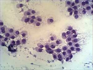
Clinical presentation: a 27-year-old man was referred for an evaluation of thyroid nodule discovered on his neck ultrasonography.
Palpation: the thyroids were not palpable.
Functional state: euthyroidism.
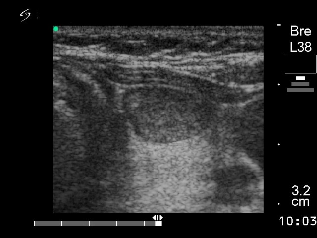
Ultrasonography: a solitary hypoechogenic nodule in the left lobe.
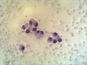
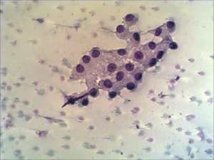
Cytological report: follicular tumor.
Histopathology: revealed a benign hyperplastic nodule.
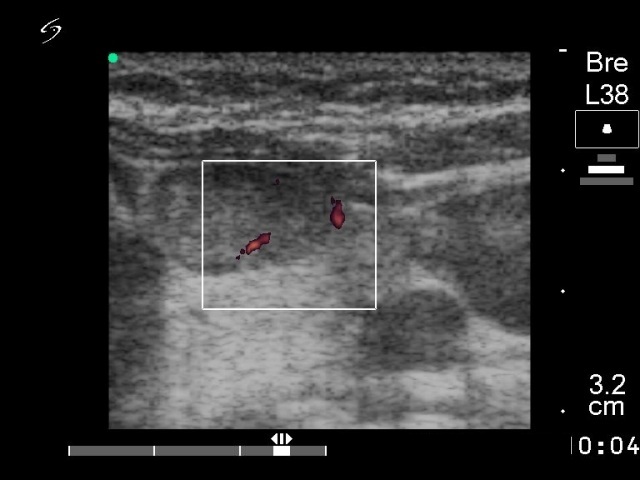
Comment: taking the cytologic and sonographic picture together, we had the chance to avoid unnecessary surgery. A follicular tumor must be surrounded with capsule. The fact that this nodule did not exhibit neither halo sign nor perinodular blood flow, significantly decreased the chance being the lesion a follicular tumor. Moreover, there was no significant atypia on the smear. Based on these two circumstances the risk of a follicular carcinoma is surely not greater than 1%. A regular follow-up instead of immediate surgery would be a better suggestion.