
|
|
Papillary carcinoma - Case 50.
|
|


Clinical data: a 44-year-old woman with a thyroid nodule known for years was referred for a yearly follow-up investigation. Previous FNAC was benign.
Palpation: a small nodule in the isthmic part of the thyroid.
Functional state: euthyroidism (TSH-level 2.91 mIU/L).
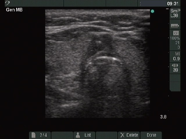
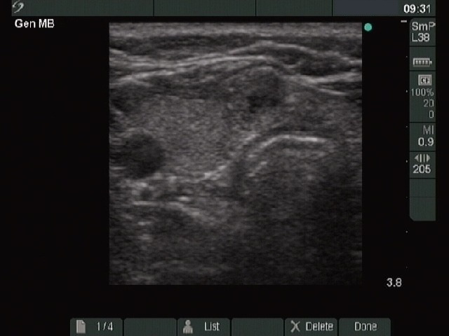
Ultrasonography: an echonormal thyroid with two hypoechogenic nodules were close to each other in the isthmus. The lesions contained microcalcification. The intranodular blood flow was not increased.
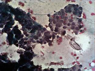
Cytological picture. There is no colloid in the background. Thyrocytes are in papillary fronds with nuclear crowding and overlapping. Many cells contain groove and nuclear inclusion.
Cytological diagnosis: papillary cancer.
Histopathology: papillary cancer.
Comment: we reevaluated the smears of previous FNAC interpreted as benign. There were only a few cells on the smear. The failure was an error of interpretation as it was in fact a not diagnostic picture.