
|
|
Papillary carcinoma - Case 49.
|
|


Clinical data: a 49-year-old man was referred for an evaluation of enlarged lymph nodes detected by himself.
Palpation: multiple enlarged firm nodules in the left side of the neck. There was no abnormality in the thyroid.
Functional state: euthyroidism (TSH-level 1.08 mIU/L).
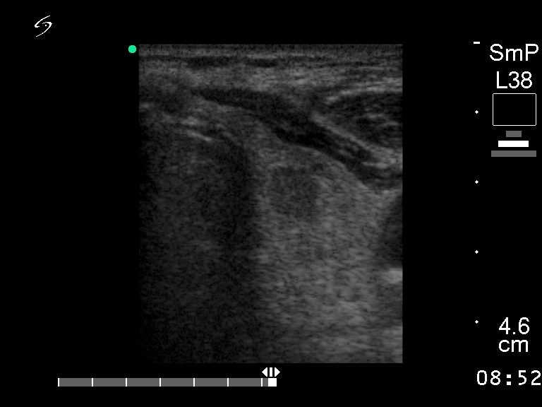
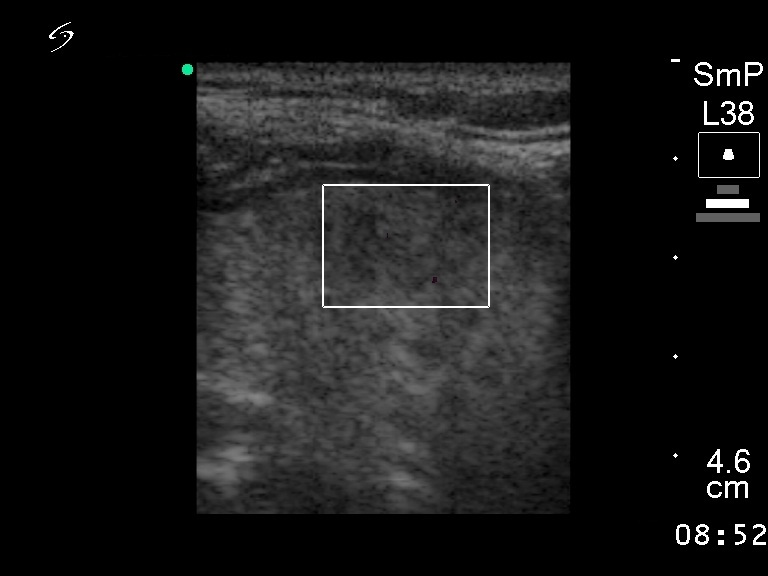
Ultrasonography: multiple enlarged lymph nodes in the left side of the neck. There was a moderately hypoechogenic lesion in the left thyroid with the dimensions of 9x7x11 mm. The lesion was avascular on Doppler mode.
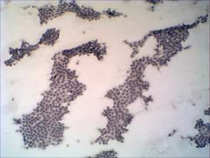
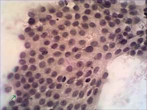
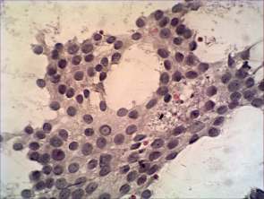
Cytological picture: no colloid in the background. Thyrocytes are located in regular monolayered regular sheets, a few of them with fibrovascular core. Follicular cells present prominent nucleoli, relatively great proportion of them exhibit nuclear inclusion.
Cytological diagnosis: suspicion of papillary carcinoma.
Histopathology: papillary carcinoma with metastasis to the regional lymph nodes and also to the lung.
Comment: the cytological picture is unusual because the clusters of follicular cells are regular, neither crowding nor overlapping of the cells can be observed. This is a rare finding in the case of a papillary cancer.