
|
|
Papillary carcinoma - Case 48.
|
|


Clinical data: A 38-year-old men was referred for an evaluation of a nodular goiter discovered by chance on routine examination. The cytology performed in another institute was benign. One year later the patient had complaints suggesting hyperthyroidism. She requested a second opinion.
Palpation: a nodule in the left lobe.
Functional state: euthyroidism (TSH 1.56 mIU/L, FT4 14.3 pM/L).
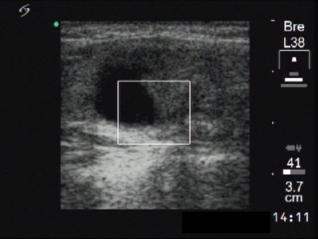
Ultrasonography: There was a mixed, cystic, moderately hypoechogenic nodule with microcalcification in the left thyroid. The lesion was avascular on Doppler mode.
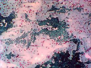
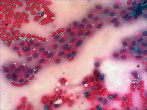
Cytological picture: There was colloid in the background. Large and small papillary clusters were observed which showed nuclear crowding. Many cells exhibit oxyphilic metaplasia. Note the presence of nuclear inclusions and grooves.
Cytological diagnosis: papillary cancer.
Histopathology: papillary cancer.