
Other edifying cases - Case 5. (ultrasonographic picture 1)
A muscle fiber mimicking a thyroid nodule

|
|
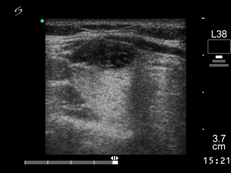
Right lobe, horizontal scan. There is a hypoechogenic lesion within or next to to the ventral part of the lobe.