
Other edifying cases - Case 5. (ultrasonographic picture 2)
A muscle fiber mimicking a thyroid nodule
|
|

|
|
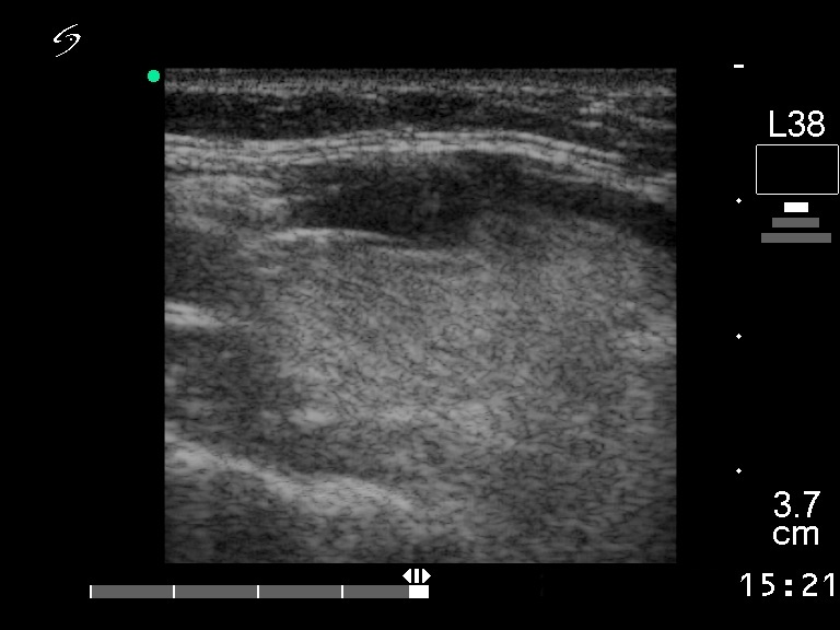
Right lobe, longitudinal scan. The hypoechogenic mass runs along the ventral surface of the lobe.