
|
|
Other edifying cases - Case 5: A muscle fiber mimicking a thyroid nodule
|
|

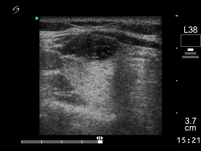
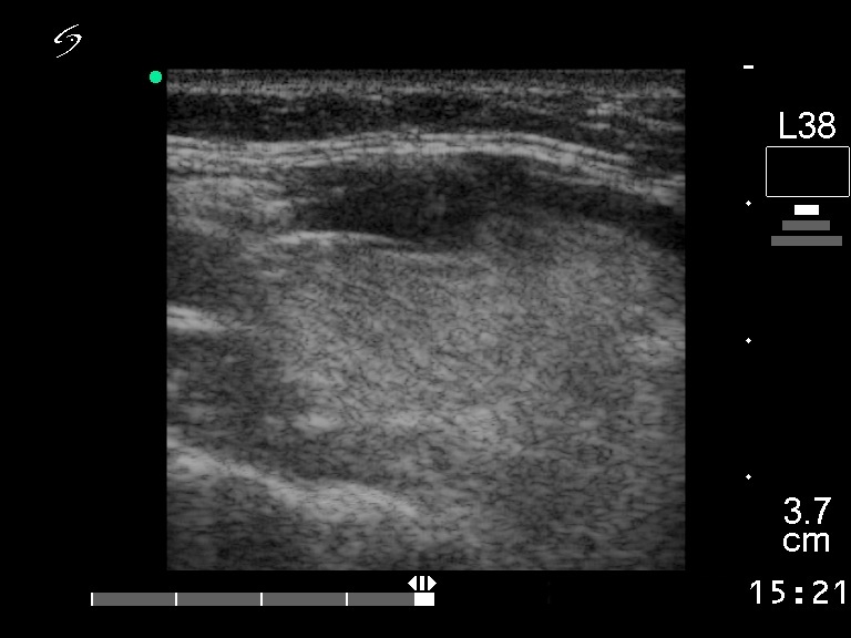
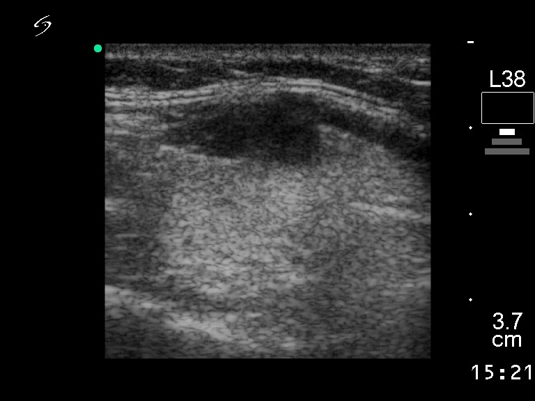
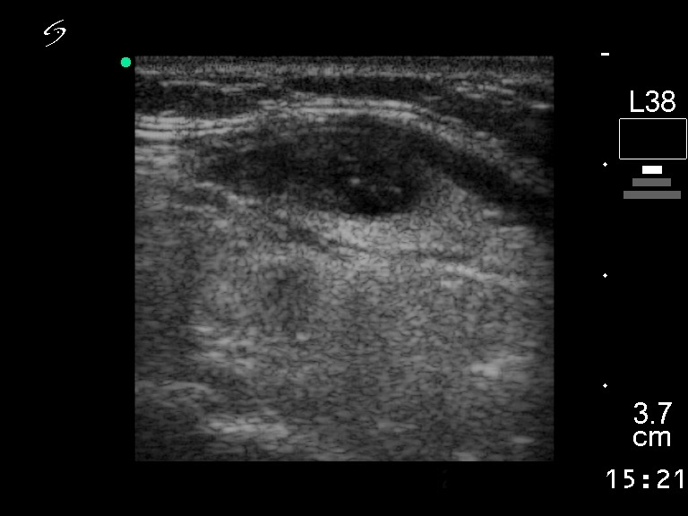
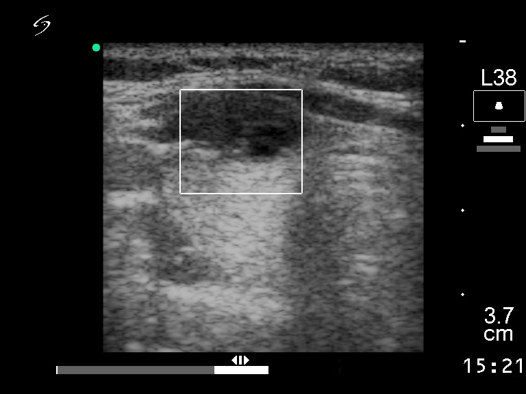
Clinical presentation: a 21-year-old woman was referred for an evaluation of a thyroid nodule detected on ultrasonography. She was examined because of upper airways infection lasting for 4 months.
Palpation: no abnormality.
Functional state: euthyroidism.
Ultrasonography: the thyroid was intact. There was a hypoechogenic mass next to the ventral surface of the right thyroid. The lesion resembled a nodule on horizontal section, while it was clear analyzing the longitudinal scan that the lesion is outside the thyroid and is a muscle fiber with great probability.