
Papillary carcinoma - Case 63. (ultrasonographic picture 3)
|
|

|
|
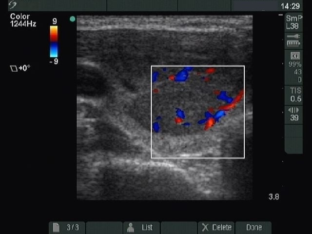
Right lobe, longitudinal scan, color Doppler method. Marked perinodular flow with increased intranodular vascularization.