Benign nodular hyperplasia - Case 30. (ultrasonographic picture 2)
|
|

|
|
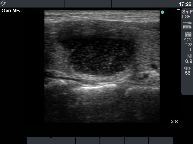
Right lobe, longitudinal scan. There is a smaller hypoechogenic lesion ventral to the larger nodule.

|
|
|
|
Right lobe, longitudinal scan. There is a smaller hypoechogenic lesion ventral to the larger nodule.