
Benign nodular hyperplasia - Case 30. (ultrasonographic picture 3)
|
|

|
|
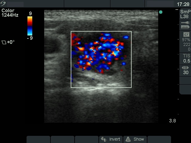
Right lobe, horizontal scan, color Doppler method. Marked intranodular flow can be seen. In this case, the cystic fluid and not vessels are responsible for the flow. Nevertheless, this is a very unusual situation.