
Benign nodular hyperplasia - Case 26. (ultrasonographic picture 1)

|
|
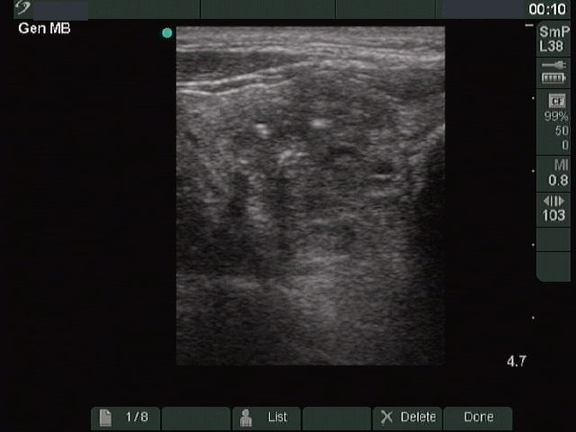
Right lobe horizontal scan. A moderately hypoechogenic nodule with irregular, not geometric border. There are three hyperechogenic granules within the nodule, the dorsal one corresponds to coarse calcification.