
Benign nodular hyperplasia - Case 11. (cytologic picture 3)

|

|

|

|
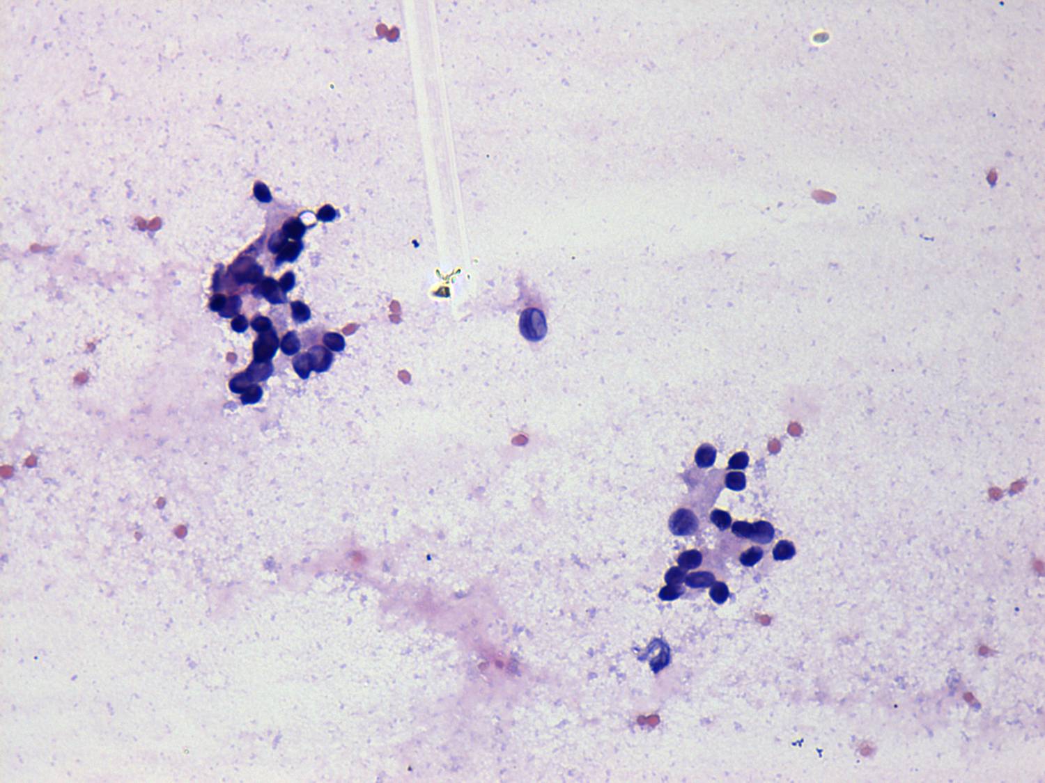
Pap-smear, 400x. The dispersed cell in the central part of the image contains inclusion.


