
|
|
Benign nodular hyperplasia - Case 11.
|
|


Clinical presentation: A 29-year-old woman was referred for evaluation of a nodular goiter detected by herself.
Palpation: an elastic nodule in the left thyroid.
Functional state: euthyroidism with TSH-level 2.09 mIU/L.
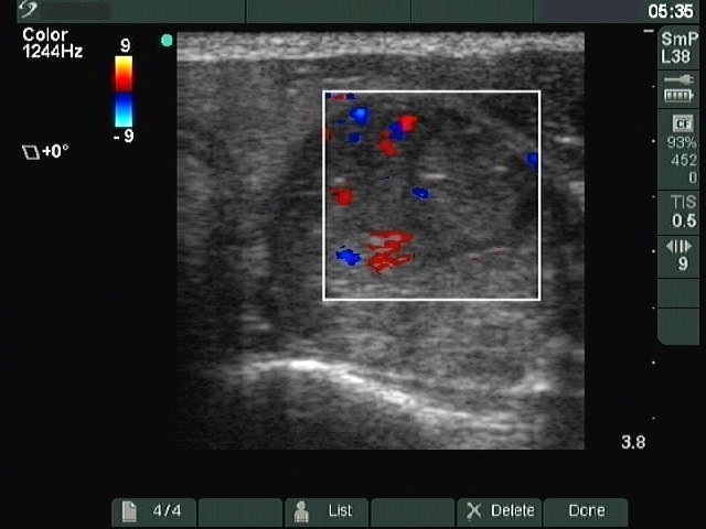
Ultrasonography: The left lobe contained a mixed nodule with increased intranodular blood flow.
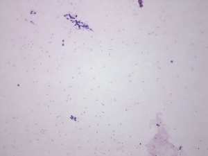
Cytology: 10 mL brown fluid was aspirated, thereafter the solid part of the nodule was punctured. Only a few cell fragments were found on the smear. Thyrocytes exhibited degenerative changes. Great proportion of the nuclei contained groove and inclusion, but it lacked other cytological signs of papillary cancer.
Cytological diagnosis: suspicion of papillary cancer.
Histopathology: benign, hyperplastic nodular goiter.