
|
|
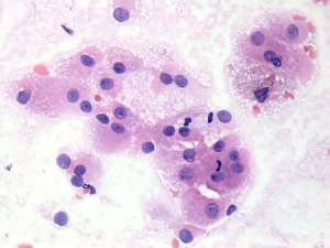
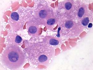
Benign nodular hyperplasia - Case 10.
|
|


Clinical presentation: a 67-year-old woman referred for evaluation of weight loss.
Functional state: euthyroidism (TSH 3.08 mIU/L, FT4 14.4 pM/L).
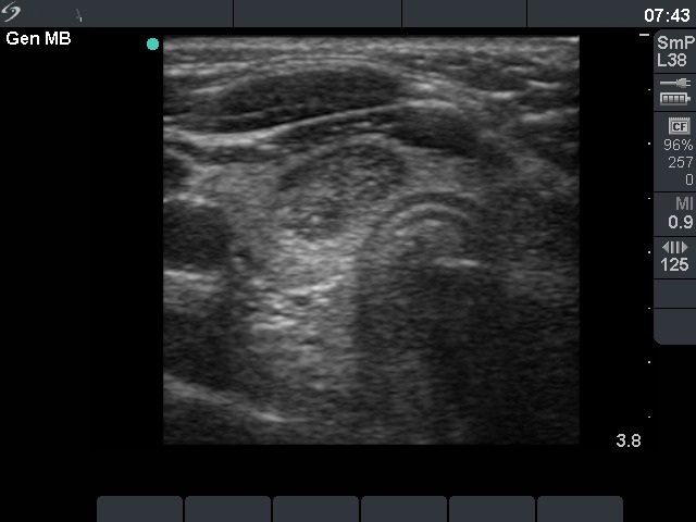
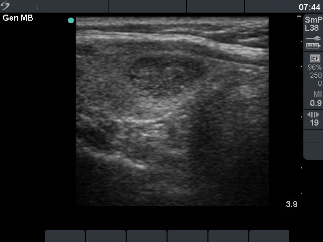
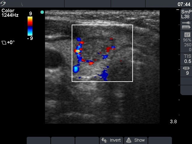
Ultrasonography: a hypoechogenic, inhomogeneous nodule with microcalcifications. The nodule did not display halo sign while did perinodular blood flow.
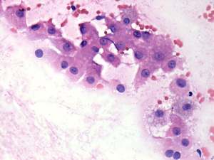
Cytology: there was no colloid in the background. Follicular cells displayed Hürthle-cell metaplasia and prominent nucleoli. Great proportion of nuclei contained groove, a few cells did inclusion. Follicular cells occurred in loose clusters and dissociated. Cytological diagnosis : Hürthle-cell tumor.
Histopathology: benign hyperplastic nodule and chronic lymphocytic thyroiditis.
Comment. This case illustrates the limitations of the preoperative diagnosis. Sonographic pattern argued for a follicular-type tumor while cytology did for a Hürthle-cell tumor. No colloid, extensive oxyphilic metaplasia, tendency of cells to dissociate, prominent nucleoli - this case had to be an oxyphilic tumor. Unfortunately, the nature does not read textbooks... Nevertheless, our task in the evaluation of patient is not to give a histopathological diagnosis.