Benign nodular hyperplasia - Case 10. (ultrasonographic picture 3)
|
|

|
|
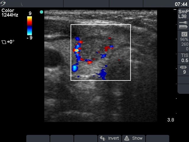
Right lobe, horizontal scan, color Doppler method. Signs of perinodular blood flow.

|
|
|
|
Right lobe, horizontal scan, color Doppler method. Signs of perinodular blood flow.