Benign nodular hyperplasia - Case 10. (ultrasonographic picture 1)

|
|
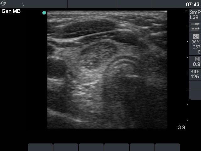
Right lobe, horizontal scan. There is a moderately hypoechogenic nodule with microcalcifications.

|
|
|
Right lobe, horizontal scan. There is a moderately hypoechogenic nodule with microcalcifications.