
Benign nodular hyperplasia - Case 21.
Initial investigation (ultrasonographic picture 1)

|
|
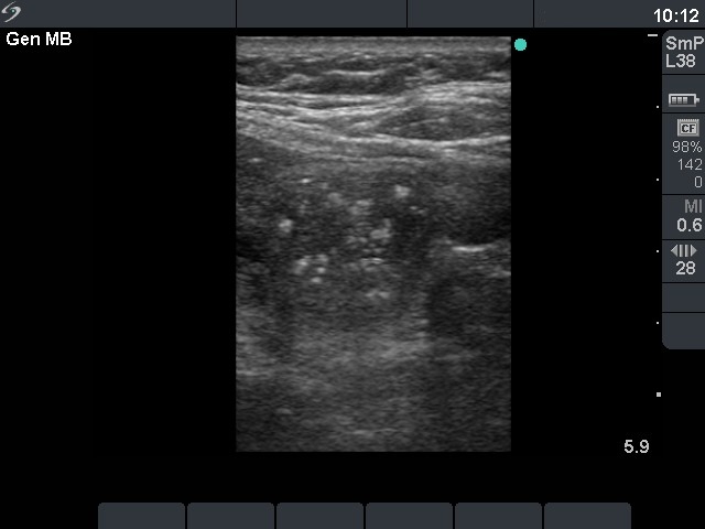
Left lobe, horizontal scan. There is a moderately hypoechogenic nodule with echonormal patches. The latter contains smaller and larger hyperechogenic granules. This pattern is practically identical to that seen in medullary cancer.